Chapter 2 - Introduction
Note
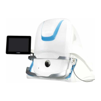
The device also includes a user guide, power cable and Cat 6 cable not shown on this diagram.
A table may also be supplied.
2.6.2 Image modalities
Optos multi-modal ultra-widefield retinal imaging enables the capture of posterior images, with a field of view of
up to 200 degrees in a single capture, and in an imaging time of less than 0.4 seconds.
Depending on the configuration of your device the following image modalities may be available:
2.6.2.1 SLO imaging modalities
optomap
optomap is an ultra-widefield (UWF
1
) color fundus image.
optomap combines neurosensory retinal and choroidal reflectance images and provides UWF image views from
a single capture action:
l
optomap color view – an UWF color fundus image combining red and green reflectance images.
l
optomap neurosensory retinal view – an UWF green laser reflectance image of the surface retina.
l
optomap choroidal view – an UWF red laser reflectance image of the deeper retina.
optomap af
optomap af is an UWF green laser auto fluorescence image.
optomap plus
l
optomap plus is a higher resolution option which is available for optomap color reflectance and
optomap af modalities.
l
optomap plus is captured at resolution x1.5 that of the standard resolution optomap.
2.6.2.2 OCT imaging modalities
CAUTION
OCT scans are described using nominal 'mm' dimensions. These dimensions are based
on an external angle scale factor of 3.47 as derived from the wide-angle schematic eye
model published by Navarro et al
2.
Optos OCT line scan
The scanner moves back and forth along the line of a selected location on the retina (fundus) and generates a
cross-sectional OCT image. The line scan is 12mm in length.
The Optos OCT line scan mode lets you capture cross sectional B-Scan OCT images of the vitreo-retina, retina
and choroid-retina structures.
This scan is also known as a B-scan OCT.
1.
Ultra-widefield images provide a field of view of up to 200° in a single capture.
2.
Isabel Escudero-Sanz and Rafael Navarro, "Off-axis aberrations of a wide-angle schematic eye model," J. Opt. Soc. Am. A 16, 1881-1891 (1999).
Page 24 of 75 Part Number: G108707/8GME
English Copyright 2018, Optos plc. All rights reserved.