Chapter 2 - Introduction
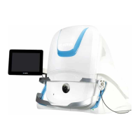
Optos OCT rnfl scan
Captures a 3.4mm diameter ring scan centered around the optic nerve head.
Example circle scan pattern
2.6.2.3 Multi-mode workflow
The device provides a multi-modal workflow sequencing option. Multi-mode assists the user in performing a
sequence of SLO and OCT scans, see Capture using Multi-mode on page51.
2.6.3 System software
The system contains features to help you configure the system and to capture, review, archive and retrieve
images. The system software comprises:
l Scan head software that enables capture of patient studies.
l Image server 'middleware' that encodes patient studies for storage.
l Image server 'archive and review' software provided by OptosAdvance.
l Browser-based review clients.
The image capture software runs on the scan head.
2.6.3.1 Understanding the review software
OptosAdvance is browser-based review software that runs on the image server. It enables eye care
professionals to manage and display the following data and images:
l Patient data
l Diagnostic data
l Images from computerized diagnostic instruments
l Images from video documentation systems
With OptosAdvance practitioners can easily view and share patient records and digital ocular scans. Detailed
instructions can be found in the OptosAdvance help files.
Physicians can use OptosAdvance to view and manage their diagnostic data, videos and images. This includes
Ophthalmic Photography, Ophthalmic Tomography, Visual Field and other relevant eye care modalities.
OptosAdvance software is used to:
l Manage and display patient data
l Store and review images and diagnostic data
You can run the review software from the following:
l
Double-click the desktop icon
Page 26 of 75 Part Number: G108707/8GME
English Copyright 2018, Optos plc. All rights reserved.