Chapter 3 - How to...
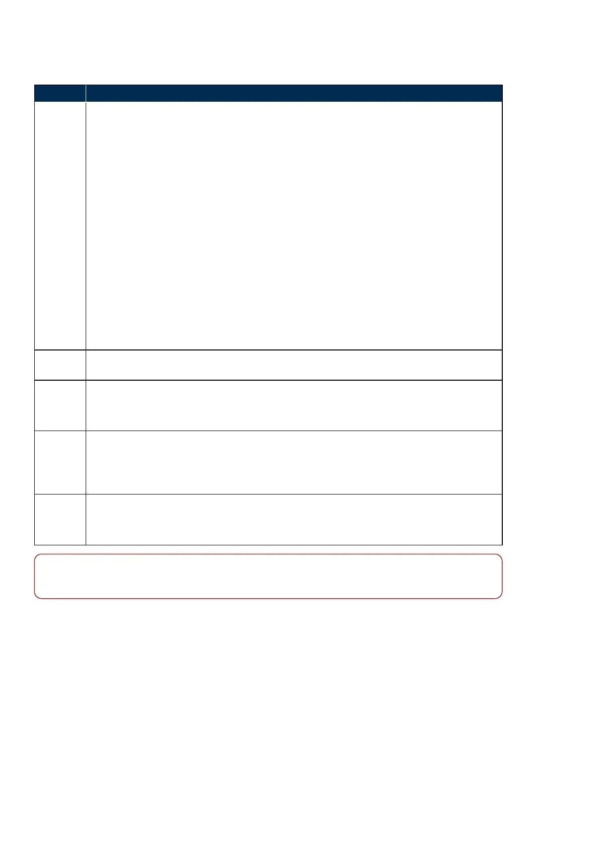
TABLE 4: Scan results
Result Description
SNR
The SNR indicator is displayed during and after the OCT image is captured.
The SNR level has a range of 10/10 (maximum) to 1/10.
The SNR level of 9/10 to 10/10 is the optimal level of SNR, where the OCT image is of the
highest quality. However, OCT SNR is impacted by the following factors:
l Patient dependent corneal birefringence
l Patient eye polarization
l Pupil size
l Voluntary and involuntary eye movements
l Misalignment of the equipment by the operator in relationship to the eye/pupil
l Poor focus on the area of interest.
To generate optimal OCT scan quality from the retina, you should attempt to capture the OCT
image at the highest possible SNR level from the captured image.
A SNR level of 5/10 is a marginal level of SNR. In this case you should repeat the scan trying to
achieve a higher level of SNR.
You should recapture the image if the SNR level is 4/10 or less.
Averages
Where image averaging is applied the scan result shows the number of scans included. For
example "Averages: 20/25" indicates 20 scans were included from 25 captured.
Movement
Movement detection indicates the extent of eye movement during capture. For example, 12/97
indicates movement detected on 12 scan lines out of the 97 captured.
You should replay the frames and assess if the movement is significant.
Blink
Blink detection indicates the number of frames where the tracking system could not detect
retinal content. For example, 18/65 indicates movement detected on 18 scan lines out of the 65
captured.
You should replay the frames and assess if this is significant.
Scan
position
error
Scan position errors indicate that the scan position cross reference to the optomap image could
not be established.
You should attempt to recapture the scan.
Note
Ocular opacity has the same impact on other retinal imaging devices as opacities block the transmission of
light and view of the retina which can then impact the SNR.
3.2.8.7 Manage the patient session
Before finishing the patient session you should check the required images and scans have been captured.
Image Browser
The Image Browser can be accessed at the end of each image capture. You can check the images and scans
which will be saved at the end of the session. You can change laterality and delete images and scans.
You can review stereo paired images. Continue capturing images until you have a good stereo pair, deleting
and recapturing as necessary.
Page 50 of 75 Part Number: G108707/8GME
English Copyright 2018, Optos plc. All rights reserved.