TEE Transducers
HD11 XE Getting Started
4535 612 62651
10
212
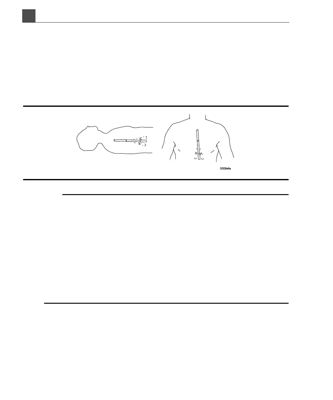
Deflection Control Basics
The deflection controls on the TEE transducers discussed in this manual move the
deflection area, located between the distal tip and flexible shaft. The deflection
area bends when you operate the controls, permitting anterior, posterior, and lat-
eral positioning for most transducers. Figure 10-2 provides an illustration of
deflection control movement.
Figure 10-2 Deflection Control Movement
ARNINGS
To prevent tissue damage such as pressure necrosis, gastroesophageal lacerations,
bleeding, tearing of adhesions, ligament damage, and perforation:
• Never apply excessive force when inserting or withdrawing the transducer, or
when operating the deflection controls.
1, 2
• Lock medial/lateral movement during insertion. Use freewheeling mode when
withdrawing the transducer any time you are not imaging.
• Never apply excessive force when operating the deflection controls during
imaging. Do not allow the transducer to remain at a maximum deflection for
long periods of time.
1, 2
1. Urbanowitz, John H. et al. “Transesophageal Echocardiography and Its Potential for Esoph-
ageal Damage.” Anesthesiology, Vol. 72, No. 1, 1990.
2. Radwin, Martin et al. “Transesophageal Echocardiography: Intubation Techniques.” Philips Appli-
cation Note 5091-2804E, 1992.