Chapter 7: Methodology and Monitoring
he EV1000 Clinical Platform NI continuously
measures the patient’s arterial pressure waveform and
calculates Cardiac Output along with other key
hemodynamic parameters. This chapter gives a brief
background on the methodology of the ClearSight
technology, instructions on how to perform a measurement
and advanced features of the system.
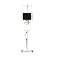
EV1000 Noninvasive System
Methodology
Accurate measurement of the patient’s blood pressure and
key hemodynamic parameters is based on the Volume Clamp
method, Physiocal method and ClearSight algorithm.
Volume Clamp Method
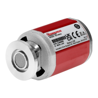
The ClearSight Finger Cuff uses the Volume Clamp method
developed by Czech physiologist J.Peñáz. The cuff is
equipped with a plethysmograph sensor, which is a combi-
nation of a light source and light receiver, to continuously
monitor changes in finger arterial blood volume. An
inflatable bladder within the cuff rapidly adjusts to this
change in volume to equilibrate the pressure of the cuff with
the pressure inside of the artery. The artery is therefore
clamped at its “un-stretched” volume and the pressure of the
cuff is equal to that of the finger arterial pressure at all times.
Physiocal Method
The Physiocal method, developed by K.H. Wesseling et al., is
short for physiological calibration. Physiocal adjusts for
changes in the “un-stretched” volume during a normal
measurement period. Cuff pressure is kept constant for one or
more heart beats and blood pressure measurement is momen-
tarily interrupted to observe the physiological properties of
the finger artery. Early in the measurement period, these
interruptions occur regularly. If the properties of the artery are
sufficiently constant over time, the interval between
Physiocals will be increased up to 70 heart beats, with higher
intervals representing increased measurement stability.
Waveform Reconstruction and Hemodynamic
Analysis (ClearSight Algorithm)
The arterial blood pressure waveform is known to gradually
change between the brachial and finger arteries due to physi-
ological reasons. The ClearSight algorithm uses advanced
processing methods to reconstruct the brachial arterial
pressure waveform (P. Gizdulich et al. 1997). Waveform
reconstruction yields beat-to-beat values of Systolic (SYS),
Diastolic (DIA) and Mean Arterial (MAP) Pressures and is
displayed.Waveform hemodynamic analysis yields values for
Cardiac Output (CO), Cardiac Index (CI), Stroke Volume
(SV), Stroke Volume Index (SVI), and Pulse Rate (PR) using
a pulse contour method (ClearSight algorithm). Advanced
algorithms are used to compute Stroke Volume Variation
(SVV) to evaluate dynamic fluid responsiveness. Systemic
Vascular Resistance (SVR), and Systemic Vascular Resis-
tance Index (SVRI) are available when a Central Venous
Pressure (CVP) value is entered.
Heart Reference Sensor
The Heart Reference Sensor (HRS) takes into account differ-
ences in pressure between the finger and heart. The hydro-
static pressure changes due to difference in height between
the finger and heart are compensated by the HRS. One ending
of the HRS is placed on the finger at the cuff level, and the
other ending is placed at heart level.
Discoloration, Numbness, or Tingling of the Fingertip
The Volume Clamp methodology places a continual pressure
on the finger which never fully occludes the arteries, but
inhibits venous return and causes some venous congestion in
the fingertip distal to the cuff. As a result, the patient's
fingertip may often experience discoloration (blue or red
coloring) after a few minutes of monitoring. After longer
periods of monitoring (approximately 30 minutes - 2 hours),
some patients may experience some tactile sensations
(tingling or numbness) in the fingertip. Immediately after
removing the cuff, the middle phalanx often shows a slightly
decreased volume and may show some reactive hyperemia or
swelling. All of these phenomena generally subside within a
few minutes of relieving the cuff pressure. Keeping the
fingers and hand warm during the measurement improves the
arterialization of the fingertip, which can improve coloration
and reduce the rate of occurrence of tactile numbing.
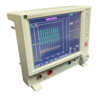
Figure 7-1 Physiocal During Blood Pressure
Measurement