Before Using this Product
42
Operation Manual
1.5 Principle of Operation
1.5.1 X-ray generating mechanism
An X-ray beam is generated using part of the kinetic energy converted when electrons rapidly
spinning decelerate in the material. To accelerate the thermoelectrons of the X-ray beam
emitted by an X-ray tube, several tens kilo voltages of high DC voltage is required. To this
requirement, an internal high-voltage transformer increases the voltage by several hundreds
voltage. The quality of the X-ray beam varies with the level of the voltage applied to the X-ray
tube (X-ray tube voltage). With higher X-ray tube voltage applied, a higher transmission
power of X-ray beam is generated. The X-ray dose depends on the magnitude of a current
owingthroughtheX-raytube(X-raytubecurrent).
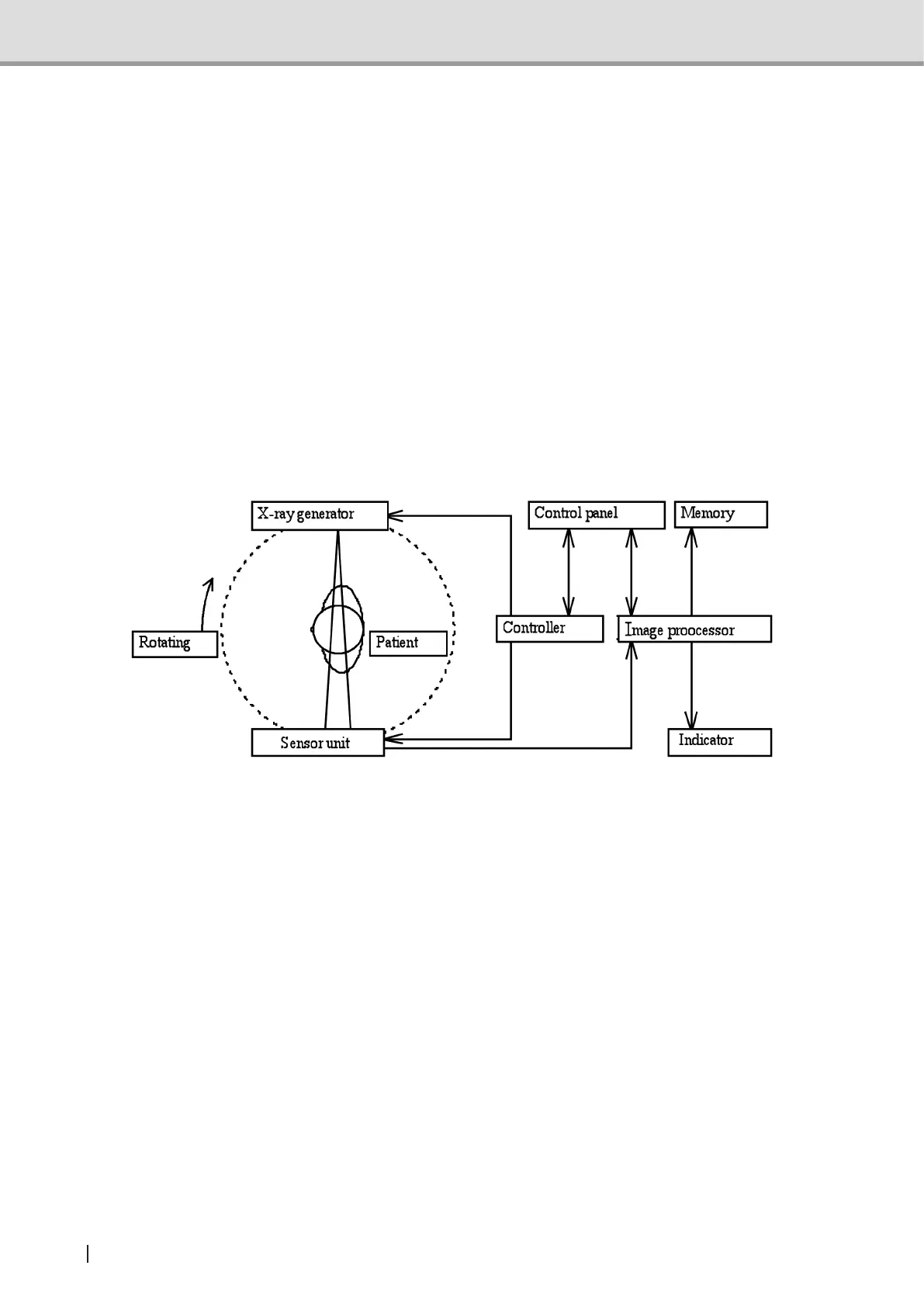
1.5.2 Operating principle
This equipment features several devices: an X-ray generator, a Sensor unit , Arm unit, which
is attached to a sliding body unit on a Column unit. While rotating around the patient's teeth
and jaw, the equipment irradiates X-ray and detects X-ray absorbed data at the Sensor unit
multiple times. Detected multiple pictures are transferred to an image processing unit and the
data is superimposed with appropriate shift value according to the X-ray moving speed by the
arm rotation to acquire panoramic image or the image of jaw joint.
To acquire Cephalometric image and Carpus image, attach the Panoramic/Cephalometric
-combined CMOS sensor to the Cephalometric unit and the equipment irradiates X-ray to head,
teeth, jaw and hand, and detect X-ray absorbed data at the Sensor unit. (*) Sync the data
transferring speed of 3D sensor and the X-ray moving speed by the arm rotation to acquire 3D
image in the image processing unit from the multiple data detected by 3D sensor.(**)
Images will be displayed on the Indicator and image data will be recorded in the memory of
magnetic disk and so on.
* For equipment with Cephalometric
** For equipment with 3D function