Probes
Vivid i User’s Manual 265
2378958-100 Rev. 02
Linear Array probes
Curved Array (Convex) probes
Doppler probe
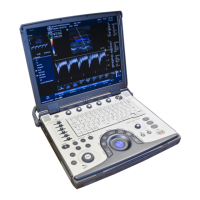
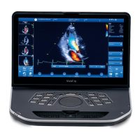
Probe Mode Intended use Technical data Image
8L-RS 2D mode
M-Mode
Color Flow
CW Doppler
PW Doppler
Peripheral
vascular
Small parts
Frequency:
Foot print:
3.2–7.5 MHz
17 x 58 mm
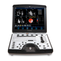
Probe Mode Intended use Technical data Image
3C-RS 2D mode
M-Mode
Color Flow
CW Doppler
PW Doppler
Abdomen
Aorto-Iliac
Fetal Heart
Obstetrics
Pelvic
Renal
Frequency:
Foot print:
FOV:
2.2–
6.0 MHz
15 x 62 mm
58 degrees
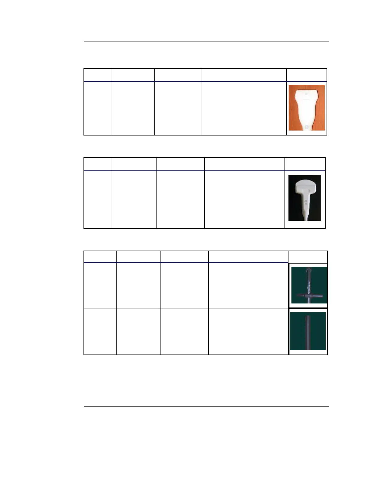
Probe Mode Intended use Technical data Image
2D
(P2D)
CW Doppler Cardiology Frequency: 2.0 MHz
6D
(P6D)
CW Doppler Vascular Frequency: 6.0 MHz
 Loading...