Special Imaging Modes
11-11
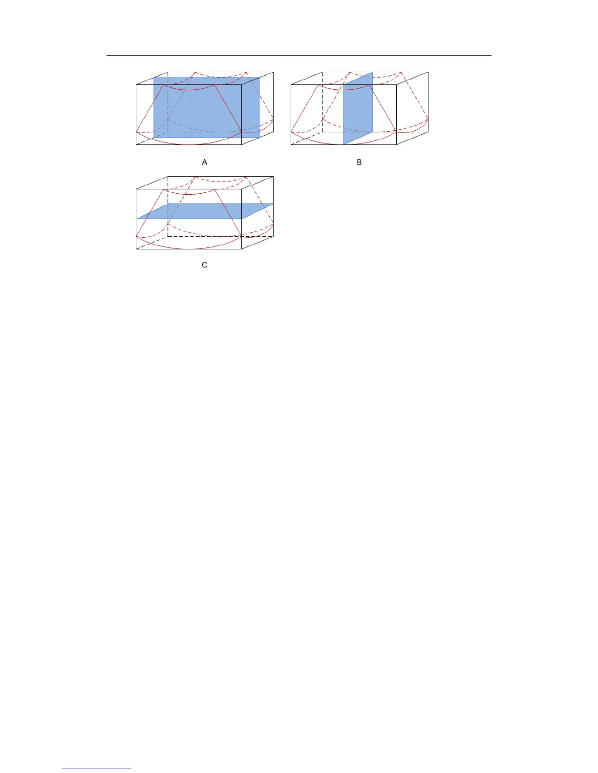
Section A: corresponds to the 2D image in B mode. Section A is the sagittal section in
fetal face up posture, as shown in the figure A above.
Section B: it is the horizontal section in fetal face up posture, as shown in the figure B
above.
Section C: it is the coronal section in fetal face up posture, as shown in the figure C
above.
Tips: the upper part of the 3D image in the D window is corresponding to the orientation
mark on the probe, if the fetal posture is head down (orientating the mother’s feet), and the
orientation mark is orientating the mother’s head, then the fetus posture is head down in
the 3D image, you can make the fetus head up by rotating the 3D image by clicking [Quick
Rot.] to be “180°” in the soft menu.
11.1.6.3 Adjust VOI (Volume of Interest)
Adjusting the VOI box size and position is to select the volume data needed to restructure
the 3D image and improve the restructure effect. The procedures are as follows:
1. In image browsing mode, click [Adjusting VOI] and set it to [On].
2. Click the [Current Window] to select a current window.
VOI on section A, B and C are adjustable.
3. Roll the track ball to adjust the VOI size, position and curve VOI position. Press <Set>
to switch among adjusting VOI size, position and curve VOI position.
To exit the VOI adjusting:
Click [Adjusting VOI] item in the soft menu to set it to [Off]. When you roll the trackball
to review the image of a section, section lines in the other two planes move
correspondingly.
Figure a and b below shows the 3D image before and after VOI adjusting.VOI adjusting
helps to restructure a better 3D image.