SSI-6000/SSI-5800/SSI-5500/SSI-5500BW
Digital Color Doppler Ultrasound System
10.1.3 Display Format
DISPLAY FORMAT
Each PW-duplex or CW-duplex mode has six possible display formats. Illustrations
for the display formats are shown in Figure 9.3, page 9-5.
10.2 PW Mode Operation
10.2.1 Sample Volume Gate Adjustment
Adjustment of the sample volume gate is possible during the real time PW mode
scan.
Follow instructions below to adjust the sample volume gate.
• Move the trackball to change the location of the gate; the spectral Doppler cursor
will be moved together. For phased array probes, the angle of the spectral Doppler
cursor is changed; for linear array probes, the spectral Doppler cursor shifts hori-
zontally.
• Press the SET key, the gate position is fixed. Move the trackball to change the gate
size.
Remark:
• Adjusting the gate position and size temporarily pause the PW spectral display
if it is active.
10.2.2 Activation of PW Spectral Display
FLOW INVERT Off
FREQUENCY 2.2
SWEEP SPEED
ECG →
BASELINE
POWER %
ANGLE CORRECT
DYN
CHROMA
VIDEO INVERT
DISPLAY FORMAT
PW↔CW
4
70
ON
8
4
2
Off
V1/2
PW
WF 225
STEER ANGLE 12
For linear array
probes only
For phased array
probes only
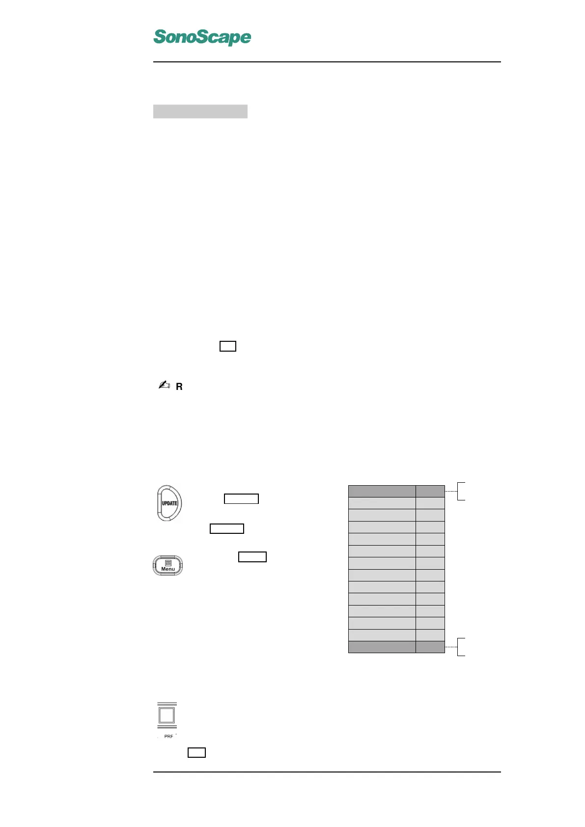
Figure 10.4: PW Mode Menu
The PW spectral is not active un-
til the UPDATE key is pressed.
With the default setting, pressing
the UPDATE key also freezes the
2D image.
Press the MENU key to activate
the PW menu (right).
10.2.3 Pulse Repetition Frequency (PRF)
The Pulse Repetition Frequency sets the velocity range of the spectral
display.
The range of the PRF depends on the probe and the application mode.
Flip the PRF switch up/down to increase/decrease the PRF range.
P/N: 4701-0061-01B
10-5
 Loading...