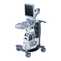
Do you have a question about the GE Voluson S6 and is the answer not in the manual?
Specifies the intended use of the system, clinical applications, patient population, and operator profile.
Provides essential safety instructions and precautions for using the ultrasound system and probes.
Outlines safety measures and maintenance requirements for the system.
Details the requirements for safely installing the system electrically, including grounding.
Provides instructions and precautions for safely moving or lifting the ultrasound system.
Covers handling precautions, watertightness, electrical hazards, and mechanical hazards related to probes.
Alerts users to potential safety hazards, such as condensation before initial use.
Step-by-step instructions for connecting power and turning on the system.
Details the procedure for safely powering off the system, including data saving.
Instructions for connecting and disconnecting transducer probes to the system.
Details the process of entering patient information for calculations and display.
Procedure for concluding an exam, saving data, and clearing temporary information.
Covers biopsy safety concerns and maintenance procedures.
Addresses specific safety concerns related to biopsy procedures and materials.
Explains the fundamental operations within the 2D mode, including gain and depth adjustments.
Details how to adjust the overall brightness of the 2D image using the gain control.
Explains how to adjust the depth range of the ultrasound image for the region of interest.
Explains how to store and review sequences of 2D images in cine memory.
Describes Contrast Imaging for enhancing flow signals and detecting abnormalities.
Details the operations and functions available within M Mode.
Explains how to adjust the overall brightness of the M mode trace.
Details MCF Mode for adding color-coded qualitative information to M mode.
Explains MTD Mode for adding color-coded qualitative information about tissue motion.
Describes MHDF Mode for incorporating flow direction into the M mode image.
Explains Doppler imaging, spectral analysis, and display elements for PW mode.
Outlines the various operations and functions available within PW mode.
Explains how to sample an area along the ultrasound beam and adjust gate size.
Details how to adjust the amplification of incoming Doppler signals for the PW spectrum.
Details how to use CW mode and adjust its settings.
Outlines the operations and functions available within CW mode.
Details Color imaging principles and how color coding provides flow information.
Outlines the operations and functions available within CF mode.
Details how Power Doppler displays slow flow velocities and its advantages.
Outlines the operations and functions available within PD mode.
Details HD-Flow Mode for incorporating flow direction into the M mode image.
Outlines the operations and functions available within HD-Flow mode.
Describes TD Mode for generating color images of tissue motion direction and velocity.
Outlines the operations and functions available within TD mode.
Details all Doppler mode adjustments, functions, and filters.
Explains how to maintain optimal resolution and accuracy from Doppler measurements.
Explains how to activate Volume mode using the 3D or 4D keys.
Steps to perform a 3D Static Volume acquisition using Sectional Planes.
Steps to perform a 3D Static Volume acquisition using Rendering.
Explains how to electronically edit images and cut away structures obstructing the view.
Explains how to save and work with acquired Volumes in 4D VolCine function.
Step-by-step guide to defining a new contour for VOCAL analysis.
Explains how to define shell contours by selecting different shell modes (OFF, Inside, Outside, Sym.).
Details on obtaining Real Time 4D mode through continuous volume acquisition and rendering.
Explains STIC for visualizing fetal heart or arteries in 4D, a post-processed 3D acquisition.
Instructions for performing Real Time 4D Biopsy, including needle track alignment.
Explains VCAD technology for automatic generation of fetal heart views for easier diagnosis.
Details how to activate the Elastography mode using the Elasto key.
Details how to use the Caliper key for generic measurements in various modes.
Explains the basic operations for generic measurements using the trackball and keys.
Lists the generic measurement methods available for 2D Imaging Mode.
Explains how to perform distance measurements like length, height, etc. in 2D mode.
Details how to measure circumference and area using trace or by setting points.
Explains how to measure volume using distances and ellipses.
Lists the generic measurement methods available for M-Mode.
Lists the generic measurement methods available for Doppler Mode.
Details how to trace the Doppler spectrum automatically to display results.
Instructions for manually tracing the Doppler spectrum to display results.
Details how to measure maximum and mean pressure gradients in Spectral-Doppler mode.
Allows measurements/calculations in 2D/3D, M, and Spectral-Doppler modes for Small Parts.
Lists distance, area, and stenosis measurements for Small Parts in 2D Mode.
Lists vessel diameter, stenosis diameter, time, and HR measurements for Small Parts in M Mode.
Lists Auto Trace, Manual Trace, and other measurements for Small Parts in Spectral-Doppler Mode.
Allows measurements/calculations for Obstetric Biometry in 2D/3D, M, and Spectral-Doppler modes.
Lists distance and area measurements for Obstetric calculations in 2D Mode.
Details how to perform distance measurements for various fetal biometry parameters.
Details how to measure circumference and area for fetal biometry using ellipses.
Instructions for calculating the Amniotic Fluid Index using multiple distances.
Explains how to calculate Nuchal Translucency (NT) for early gestation.
Lists FHR (Fetal Heart Rate) measurements for Obstetric calculations in M Mode.
Lists Auto Trace, Manual Trace, and other measurements for Obstetric calculations in Spectral-Doppler Mode.
Contains measurements to obtain Z-scores for fetal heart analysis.
Details measurement for calculating fetal limbs and estimating fetal weight.
Allows measurement of the fetal heart in 2D/3D, M, and Doppler modes.
Allows measurements/calculations for Cardiac applications in 2D/3D, M, and Spectral-Doppler modes.
Lists various measurements for LV, AV/LA, MV, TV, PV and LVOT/RVOT in 2D Mode.
Details calculation of left ventricular volume in 2D mode.
Explains calculation of Volume Area/Length in 2D mode.
Explains measurement of Left Ventricular volume and mass.
Details methods to calculate Mitral Valve measurements in M mode.
Explains how to measure Tricuspid Valve diameter in 2D mode.
Details how to measure Aortic Root Diameter and Left Atrium Diameter in 2D mode.
Explains how to measure Pulmonary Valve Diameter in 2D mode.
Lists measurements for LV, AV/LA, MV, and HR in M Mode.
Describes two methods to calculate the Left Ventricle in M mode.
Step-by-step guide to calculate all Left Ventricle items in M mode simultaneously.
Lists measurements for MV, AV, TV, PV, LVOT/RVOT Doppler in Spectral-Doppler Mode.
Allows measurements/calculations for Urology applications in 2D/3D, M, and Spectral-Doppler modes.
Lists distance, area, and stenosis measurements for Urology calculations in 2D Mode.
Lists vessel diameter, stenosis diameter, time, and HR measurements for Urology calculations in M Mode.
Lists Auto Trace, Manual Trace, and other measurements for Urology calculations in Spectral-Doppler Mode.
Allows measurements/calculations for Vascular applications in 2D/3D, M, and Spectral-Doppler modes.
Lists distance, area, and stenosis measurements for Vascular calculations in 2D Mode.
Lists vessel diameter, stenosis diameter, time, and HR measurements for Vascular calculations in M Mode.
Lists Auto Trace, Manual Trace, and other measurements for Vascular calculations in Spectral-Doppler Mode.
Allows measurements/calculations for Gynecology applications in 2D/3D, M, and Spectral-Doppler modes.
Lists distance, angular, and area measurements for Gynecology calculations in 2D Mode.
Lists vessel diameter, stenosis diameter, time, and HR measurements for Gynecology calculations in M Mode.
Lists Auto Trace, Manual Trace, and other measurements for Gynecology calculations in Spectral-Doppler Mode.
Allows measurements/calculations for Pediatrics applications in 2D/3D, M, and Spectral-Doppler modes.
Details Hip Joint measurements for Pediatric calculations in 2D Mode.
Explains the Hip Joint calculation for assessing infant hip development.
Allows measurements/calculations for Neurology applications in 2D/3D, M, and Spectral-Doppler modes.
Lists distance, area, and stenosis measurements for Neurology calculations in 2D Mode.
Lists vessel diameter, stenosis diameter, time, and HR measurements for Neurology calculations in M Mode.
Lists Auto Trace, Manual Trace, and other measurements for Neurology calculations in Spectral-Doppler Mode.
Explains how the Calculation function is switched on and caliper appears.
Explains how to set up DICOM target nodes for image transmission.
Instructions on how to enter the Measure Setup section.
Details the four different actions that can be programmed onto P-keys (P1-P4).
Instructions on activating Start Exam, including probe and application selection.
Details the configuration options for the End Exam button, including archive and review settings.
Provides guidelines for safely interconnecting medical devices and auxiliary equipment.
Crucial safety rules regarding ECG module usage, electrical hazards, and defibrillation.
Lists the safety standards and certifications the Voluson® S6/S8 conforms to.
| Power Requirements | 100-240 VAC, 50/60 Hz |
|---|---|
| Connectivity | DICOM, USB, Ethernet |
| Application | Obstetrics, Gynecology |
| Display | 15-inch LCD monitor |
| Imaging Modes | Color Doppler, Power Doppler |
| Probes | Convex, Linear, Phased Array, Endocavitary |
| Storage Capacity | Hard drive (capacity unspecified) |