ZEISS OPERATION Axio Imager 2
Illumination and contrast methods
184 430000-7544-001 01/2016
(3) Setting reflected-light brightfield according to KÖHLER
− The microscope should be set up correctly as described in Section 3.
− Switch on the microscope.
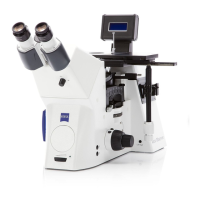
• Switch on the halogen lamp for reflected light using the reflected-light / transmitted-light toggle
switch (Fig. 76/36) on the microscope stand.
Depending on the equipment installed, the microscope contains a 6x20 compensator mount or a 4-
position modulator turret for setting the contrasting techniques. The 6x20 compensator mount can be
used for both brightfield and darkfield. For C-DIC and TIC examinations, the corresponding 6x20 slider is
also required. Refer also to Section 4.12.9.
The 4-position modulator turret has a combined brightfield/darkfield position (H/D) as well as three
additional positions for C-DIC (C1, C2) and TIC (TIC). Refer also to Section 4.12.10.
• If the 6x20 compensator mount is used, remove the 6x20 slider, if necessary. If the 4-position
modulator turret is used, set the H/D position.
• Swivel the reflector turret into brightfield position H.
• Adjust light-intensity control (Fig. 205/5) on the microscope stand.
• Place a high-contrast reflected-light specimen on the stage.
• Turn nosepiece (Fig. 205/7) to swing in 10x objective (yellow ring, see also Section 2.5).
• Use focusing drive (Fig. 205/6) to focus on the specimen. In doing so, always focus away from the
specimen, if possible, to avoid any collision between objective and specimen.
• Remove the reflected-light diffusion disk. Turn the adjusting screws of the HAL 100 halogen
illuminator to focus and center the image of the lamp filament in the exit pupil of the objective. To do
so, either pull out the adjusting aid or remove one eyepiece from the binocular tube. Afterwards, push
the adjusting aid in again or reinsert the eyepiece. Move the reflected-light diffusion disk into the light
path again.
• Set the aperture diaphragm (Fig. 205/2) in mid-position (roughly half open or closed) by turning its
knurled wheel.
• Reduce the size of the luminous-field diaphragm (Fig. 205/4) by turning its knurled wheel until it
becomes visible in the field of view (Fig. 205/A).
• Turn the focusing drive (Fig. 205/6) to refocus on the edge of the luminous-field diaphragm
(Fig. 205/B) and (using the SW 3 ball-headed screwdriver) turn the centering screws (Fig. 205/3) until
the luminous-field diaphragm is concentric with the edge of the field of view (Fig. 205/C).
• Then, open the luminous-field diaphragm (Fig. 205/4) so that it just disappears from the field of view
(Fig. 205/D).
• To set the aperture diaphragm (image contrast), remove one eyepiece from the binocular tube and
look into the tube with the naked eye or insert the auxiliary microscope in place of the eyepiece.
• Center the aperture diaphragm with the centering screws (Fig. 205/1) and, for specimens with average
contrast, adjust the size of the aperture diaphragm to about 2/3 to 4/5 of the exit pupil diameter of
the objective (Fig. 205/E) by means of knurled wheel (Fig. 205/2).
In most applications, this aperture diaphragm setting provides optimum contrast at almost ideal
resolution, and is therefore the best compromise for the human eye.
• Finally, reinsert the eyepiece, refocus with the coaxial coarse and fine focusing drive (Fig. 205/6) and
adapt the image brightness to the specimen being examined.
Never use the aperture diaphragm to control the image brightness. Instead use the light
intensity control (Fig. 205/5), or swivel attenuation filters of the 2-position filter wheels into the
light path!