6 Image Acquisition
Operator’s Manual 6 - 23
Perform the following procedure:
1. During real-time scanning, select [MFE] to start MFE imaging.
2. Use [MFE Period] to select different imaging period suitable for current flow. Where, MAX is
the maximum superimposing effect.
6.13.6 Contrast Imaging QA
Contrast Imaging QA images are provided for reference only, not for confirming
a diagnosis.
• In case of inaccuracy of the data, do not adjust the depth and the pan-zoom when saving the
cine.
• If the contrast signal inside the selected ROI does not meet the requirements of gamma fitting
condition, that is the bulleting injection, curve fitting may not be available.
Contrast Imaging QA adopts time-intensity analysis to obtain perfusion quantification information
of velocity flow. This is usually performed on both suspected tissue and normal tissue to get
specific information of the suspected tissue.
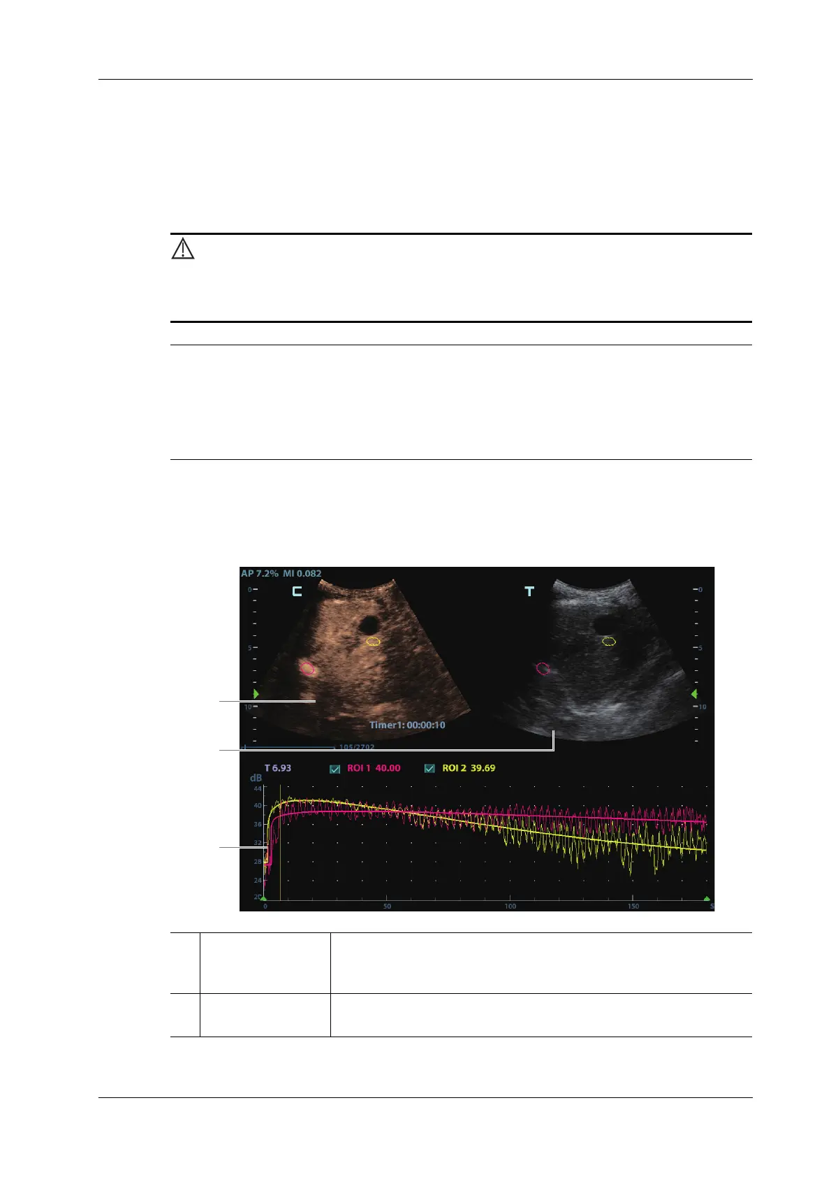
Figure 6-1 Contrast QA Screen
1 Contrast cineloop
window
Sample area: indicates sampling position of the analysis curve. The
sample area is color-coded, 8 (maximum) sample areas can be
indicated.
2 B cineloop window Sample areas are linked in the contrast cineloop window and B
cineloop window.