Instructions for Use 8 Operation
2660021169042 Rev. A 2018-03 8.4 Determine Scan Type
2660021169042 Rev. A 2018-03 81 / 246
PLEX® Elite 9000 Angiography uses differences between B-scans to
generate contrast associated with the motion of blood through the
vasculature. Angiography uses swept-source frequency filtering to
generate images with detailed vasculature.
To image vascular flow, each B-scan in the scan pattern is repeated
several times consecutively. Comparisons of the signals on consec-
utive B-scans in the same location reveal some areas with signal
change over time and some areas with constant signal. Temporal
signal change in a specific location is considered erythrocyte
motion and indicates a vessel location.
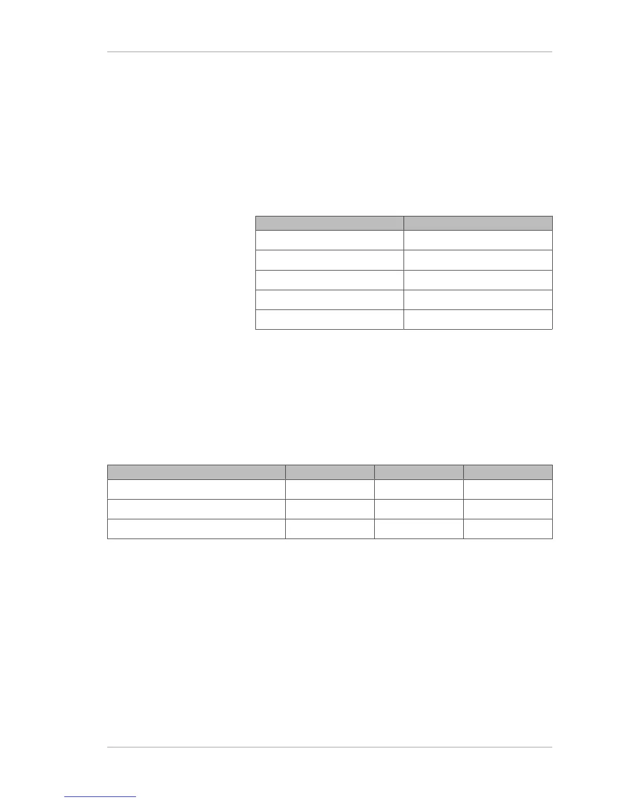
Scan Name Area
Angio (3mmx3mm) 3 x 3 mm
Angio (6mmx6mm) 6 x 6 mm
Angio (9mmx9mm) 9 x 9 mm
Angio (12mmx12mm) 12 x 12 mm
Angio (15mmx9mm) 15 x 9 mm
Table10: Angio Scans
HiRes Tracking is available (and turned on) for Angio
(3mmx3mm) . You can turn off this feature.
8.4.1.2 Cube Scans
PLEX® Elite 9000 Cube scans combine A-scans to generate a series
of B-scans and a 3-D macular thickness map.
Cube scans acquire a series of horizontal scan lines to generate a
cube of data through a grid.
Scan Name Grid Size # of Scan Lines # of A-scans
Cube (12mmx12mm) (1024x1024) 12x12 mm 1024 1024
Cube (12mmx12mm) (800x800) 12x12 mm 800 800
Cube (12mmx12mm) (512x512) 12x12 mm 512 512
Table11: Cube Scans
8.4.1.3 HD Scans
Two types of HD Scans are available:
• Spotlight 1 (16mm) (10-100x)
Spotlight 1 (16mm) (10-100x) scan generates a single, high-
definition scan with the following features:
– Depth: 3.0 mm
– B-scans:100
– A-scans:1024
– Length: 16 mm (adjustable to 4mm, 6mm, 9mm, 12mm)
 Loading...