2 Introduction Instructions for Use
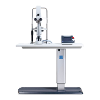
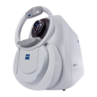
2660021169042 Rev. A 2018-032.6 PLEX® Elite 9000 Technology
34 / 246 2660021169042 Rev. A 2018-03
In Swept Source OCT, spectral data is acquired over time by illumi-
nating the interferometer with a light source that produces only a
very narrow optical frequency band at a given moment, and then
rapidly shifts that band across a broad spectrum. A fast detection
mechanism observes the output of the interferometer, and the
interference spectrum over the broad band is measured. Swept
Source OCT imaging of the retina can be performed using light
with a wavelength near 1060 nm, a wavelength longer than the
840 nm used in Stratus™ OCT and CIRRUS™ HD-OCT. Clinical
reports have consistently shown a measureable improvement in the
imaging performance in deeper tissues, particularly beyond the
retinal pigment epithelium (RPE). There are a number of possible
reasons for this increase in penetration, including reductions in
scattering and absorption in some tissues. Reduced absorption by
melanin in the RPE not only allows light to pass more easily to the
tissues beneath, but reduces the potential for localized heating due
to laser exposure.
Swept Source OCT has several practical advantages over SD-OCT
(Spectral Domain OCT). Swept source implementations exist today
that make it possible to resolve the spectrum at much higher
resolution than can be achieved with a spectrometer array at
reasonable cost, which translates to both an increase in the
possible depth window that can be simultaneously displayed and
less signal roll-off across that depth. Swept sources are also
associated with OCT maximum speed, although this difference is
not fundamental. Finally, Swept Source OCT is fundamentally less
sensitive to motion-related signal loss.
2.6.1 New in Version 1.7
2.6.1.1 High-Definition B-scans (HD-51 Lines)
A new scan option, HD 51 Line, consists of 51 B-scans evenly
distributed over the FOV. You can select the number of repeated B-
scans: 5, 10, 15 or 20.
HD 51 Line scans are available for the following scans:
• Cube 6mmx6mm , 5, 10, 15, or 20 repeated B-Scans
• Cube 12mmx12mm , 5, 10, 15, or 20 repeated B-Scans
non HD HD 51 Line
Table1: Cube 12mmx12mm B-Scan Examples