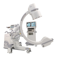
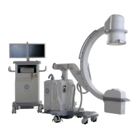
Chapter 7: Digital Detector
5495975-1EN Rev.9 7-1
© 2013-2017 General Electric Company. All rights reserved.
Chapter 7: Digital Detector
This section outlines the basic detector functions, usage, care, and specifications.
Detector Overview
Detector primary functions are:
To convert x-ray data into digital image data.
To transfer the digital data to an external workstation for processing and display.
The detector is an x-ray imaging device. It consists of an array of 2022 x 2022 pixels (40.4 x 40.4 cm).
Each pixel is attached to a data acquisition circuit that converts incoming x-ray signal to 14-bit digital
data.
The detector is constructed from a carbon fiber. The front Surface contains a graphite x-ray imaging win-
dow. The back surface of detector contains safety warnings. Detector enclosure is identified as applied
part.
Figure 7-1 Detector Overview
Note: The back surface of the detector contains screws and should not be imaged or exposed. Place
this surface away from the patient.
Panel
The panel consists of a thin-film amorphous silicon integrated circuit on a glass substrate with a cesium
iodide scintillator. The scintillating material absorbs the x-rays and converts the energy to light. The light
is converted into a charge that is digitized by the detector electronics.
 Loading...