Chapter 3 Page no. 46
Introduction.fm
GE Healthcare Senographe DS
Revision 1 Service Information and Procedures Class A 2385072-16-8EN
Sub-Systems and Components
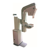
2-2. Senographe X-ray System
The Senographe is equipped with a dual track X-ray tube (molybdenum/rhodium) and a Digital Detector.
Mammographic examinations can be made with standing, sitting, or recumbent patients; both contact
and magnification views are available.
Images are acquired by direct digitization; they are displayed immediately on the LCD monitor and are
stored for later diagnostic review. They can be processed and/or filmed.
AOP (Automatic Optimization of Parameters) and manual setting modes are provided for control of the
X-ray parameters; the system provides auto-collimation.
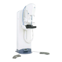
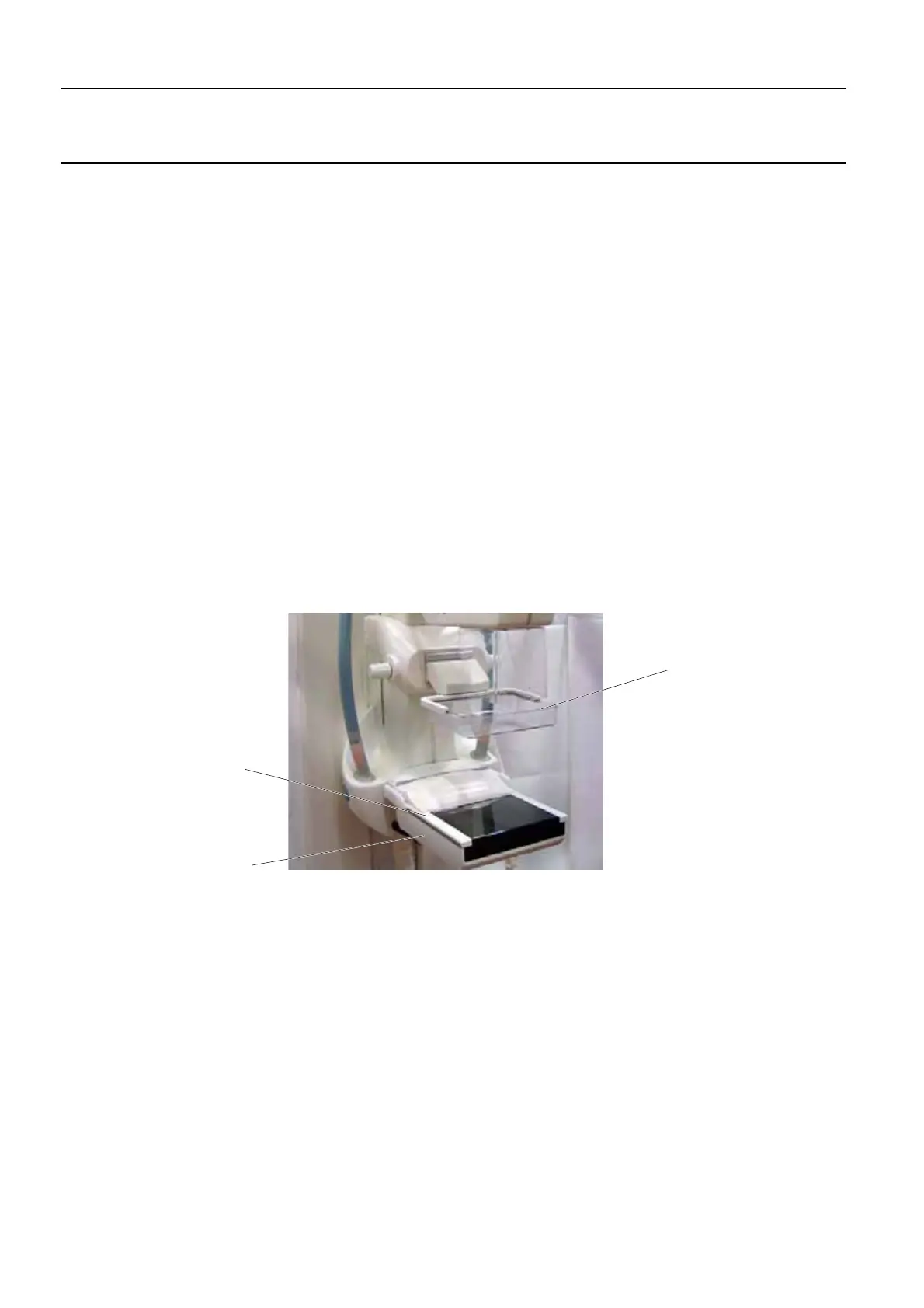
2-3. Digital Detector and Image Receptor
The Digital Detector is built into the Image Receptor, shown below. It is a flat panel of amorphous silicon
on which cesium iodide is deposited to maximize detection of X-Rays and transmission of light photons.
The high definition digital images produced are sent to the Acquisition Workstation for visualization and
processing.
A removable grid (Bucky) plugs into the Arm above the Image Receptor. For magnification views, Mag
Stands providing magnifications of 1.5 or 1.8 are used instead of the Bucky. The presence of the Bucky
or the Mag Stand is recognized automatically.
Image Receptor
Compression
Paddle
Bucky