71
Software
D
D
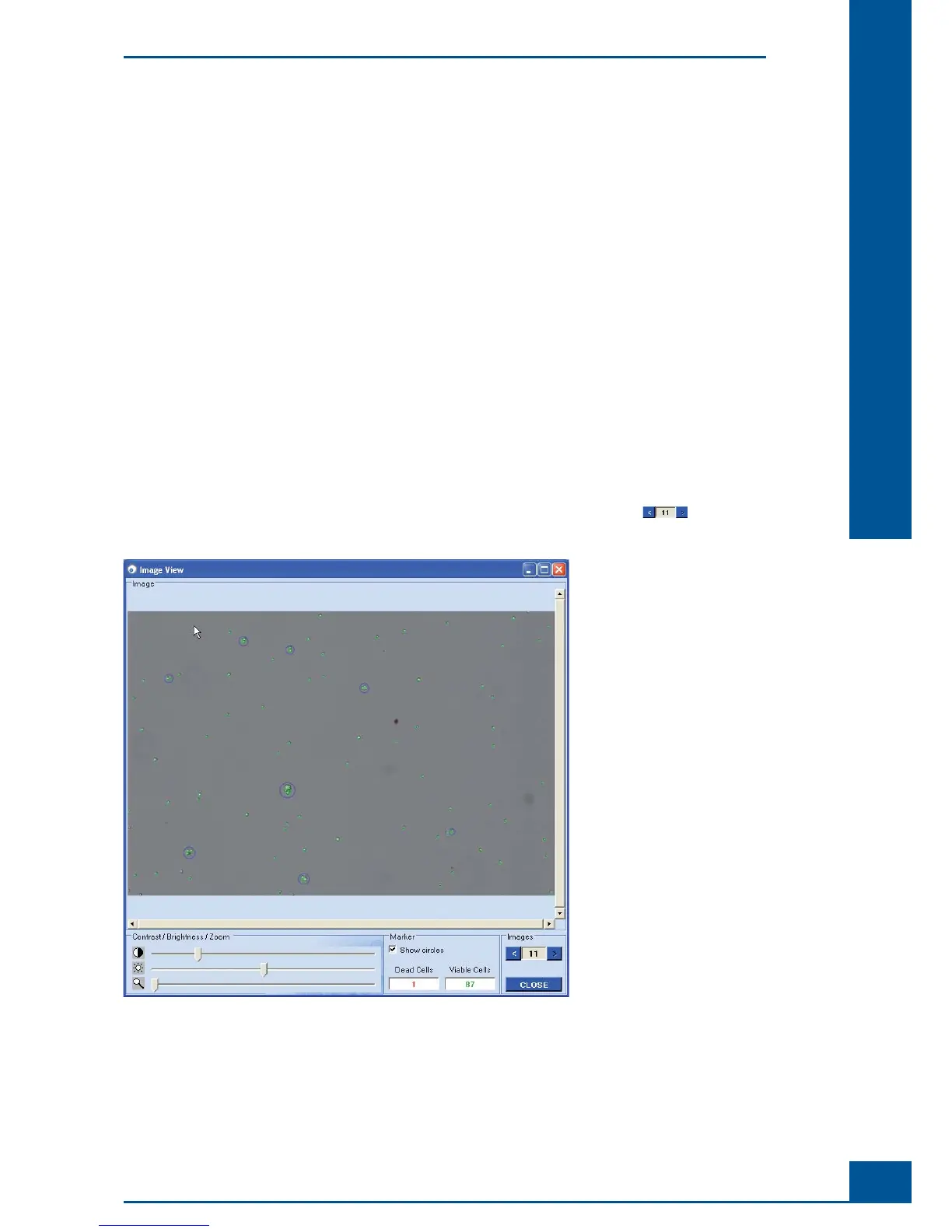
1.3.2. Viewing a Cell Image Using the Image View Window
It is possible to see which cells were marked viable and dead cells by the analysis software via the Image View
window. The user can thus get a sense of how cell shapes and clusters have been evaluated by the software.
The Cedex HiRes Software 2.2 offers various options for optimizing measurement results by adjusting the
Operator used for the image analysis. Thus, the image analysis Operator can be adjusted for abnormal cell
culture images or for specifi c internal counting strategies. For more information about adjusting the image
analysis, see “
Adjustment of the Image Analysis using the Live Operator”.
The Image View offers the following information and options:
Viable Cells: number of viable cells.
Dead Cells: number of dead cells.
Show Circles check-box: switches on/off the marking of cells recognized as living or dead. Viable cells
are marked with a uniform green circle and dead cells are marked with a red circle. The circle diameter
corresponds to the cell diameter. Objects are marked with a blue circle.
Zoom: Image enlargement.
Brightness and contrast settings.
Navigating a window when the image is enlarged (scroll bars appear at the bottom and right edge of the
image when the image is zoomed in).
Scrolling through all of the images for a particular measurement using the arrows in the lower
right-hand corner.
Figure 52: Viewing a cell image using the Image View window
Using the Measurement Results Window
Image Area