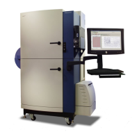
FLIPR
®
Tetra High Throughput Cellular Screening System User Guide
0112-0109 H 107
method will be labeled with the name of the data file it was created
from. However, if a single export file is desired that contains
information for multiple data files, select Use user-defined name in
the respective section. In this instance, all exported information will be
combined into one file labeled with the desired user-defined name.
When the file is open, the individual data file names which the
information was exported from are used as the header for each data set
within the export file.
Image Display
Use the Image Display dialog, opened from the Analysis process
page, to view images saved in data files when Save Images was
enabled during a Read with TF step. This option stores a total of 100
images per experiment; the number of images per dispense is
user-defined. The default is set to one image before and nineteen
images after the fluid addition is initiated. Saved *.tif files can be
played back in sequence, frame-by-frame or as a video.
Image Display is useful for diagnosing problems. For example, if cells
are blown off during a fluid dispense, you will see dark holes in the
middle of the cell layer after the fluid addition. If the entire well
decreases in relative light units, it is likely that extracellular dye has
been diluted.
Notes
In protocol files, open the Protocol Notes dialog where you can enter
comments that you want kept with all data files generated with the
protocol. The dialog contains a simple text editor for you to format your
comments.
In data files the same dialog shows the comments that were added in
the protocol file, now in read-only format, so they cannot be edited.

 Loading...