Running an Experiment
184 0112-0109 H
ion-free buffers. For example, if studying a calcium channel, dye load
cells using a calcium-free buffer and prepare your compound plate
using a calcium containing buffer.
Effect of DMSO on Membrane Potential Assays
High concentrations of DMSO are toxic to many cell lines and affect
compound mixing. Also, DMSO can induce physiological changes in cells
(for example, differentiation). If the test compound preparation
contains DMSO, it is important to titrate the DMSO to determine the
maximum concentration that can be used with each indicator cell line
and test compound. With some cell lines that have been tested, there
was no effect on signal level up to 1% DMSO final concentration.
Voltage Sensor Probes Assay Protocol
This section provides the following information and procedures:
• Materials required for the assay
• Cell preparation guidelines
• Dye loading procedure
Required Materials
The following materials are required for running a membrane potential
assay:
Item Source
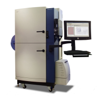
FLIPR
®
Tetra System with Voltage
Sensor Probes Optics Kit installed:
FLIPR
®
Tetra System LED Module
390–420 nm FLIPR
®
Tetra System
Emission Filter 440–480 nm FLIPR
®
Tetra System Emission Filter 565–625
nm
Molecular Devices
See Appendix C
Voltage Sensor Probes:
DisBAC2(3) and CC2-DMPE
VABSC-1 Background Suppression Dye
Invitrogen
K1016
K1019
Clear, flat-bottom, black- or clear-wall
384- or 96-well plates
See Appendix C
Clear polypropylene source plates See Appendix C
FLIPR
®
System pipette tips, 96 or 384
See Appendix C
Cells in suspension —
Test compounds Specific to receptor

 Loading...