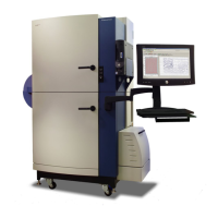
FLIPR
®
Tetra High Throughput Cellular Screening System User Guide
0112-0109 H 35
of the frames collected during the exposure time. This controls the
signal intensity of the assay.
The recommended value range for the ICCD camera is 40,000 to
50,000 (at maximum signal). This camera does not require
temperature cycling when changing between Fluorescence and
Luminescence modes.
Both Cameras
Once calculated, RLUs are displayed in real time in the ScreenWorks
Software Analysis process window (within the limits of computer
processing speed). Data for one wavelength (for example, Read
mode), for all 1536, 384 or 96 wells of the read plate, are displayed in
the Multi-Well Graph on the right side of this view. The Detail Graph
can be populated by selecting wells in the Multi-well graph. When an
experiment is completed, post-assay analysis can be done via
ScreenWorks Software, or data can be exported.
In normal operation images are discarded once RLU values have been
measured. However, for quality control purposes, users can define the
number of images per Read with Transfer Fluid step. Up to 100
images per experiment can be retained. These images can be useful for
troubleshooting problems, such as cells lifting from well bottoms during
compound addition. Images are saved as *.tif files with the same name
and to the same directory as the data file. They can be viewed by
clicking the Image button in the Analysis process page when the
resulting data file is open in ScreenWorks Software.
Please note that the robustness of an assay is not dependent on the
size of the signal; it is better determined by the signal-to-noise ratio. A
commonly used calculation for determining assay robustness is the Z’
factor equation
1
.
LED Modules
The FLIPR
®
Tetra System has a total of four LED banks providing
illumination for plate reading. The LED banks pulse on, two at a time,
only when an image is to be captured, protecting cells from possible
dye photo-bleaching.
The LED banks are divided between two modules, one on either side of
the read plate position above. LED banks in corresponding positions in
either module are paired. The paired LED banks pulse simultaneously
during an experiment, so that light strikes the read plate base from two
directions, helping to ensure that the entire plate is maximally
illuminated.
1. Zhang J, Chung TDY and Oldenburg KR. A Simple Statistical Parameter for Use
in Evaluation and Validation of High Throughput Screening Assays. J. Biomol.
Screen. 1999; 4:67-73.

 Loading...