GE HEALTHCARERAFT VOLUSON E8 / VOLUSON E6
D
IRECTION KTD102576, REVISION 7 DRAFT (AUGUST 23, 2012) SERVICE MANUAL
4-20 Section 4-4 - Functional Checks
• VCI OMNI VIEW
Volume Contrast Imaging Omni View (any plane) - improves the contrast resolution and the signal
/ noise ratio and therefore facilitates finding of diffuse lesions in organs.
Volumes can be edited in all other Visualization Modes.
• STIC The fetal heart or an artery can be visualized in 4D (also in combination with PD, HD-Flow or CFM)
• 4D BIOPSY
Real Time 4D Biopsy - continuous volume acquisition + parallel calculation of 3D rendered images
1
QUALITY
Changes the line density against the acquisition speed (low, mid1, mid2, high1, high2).
2
VOL. ANGLE
To select the Volume Sweep Angle.
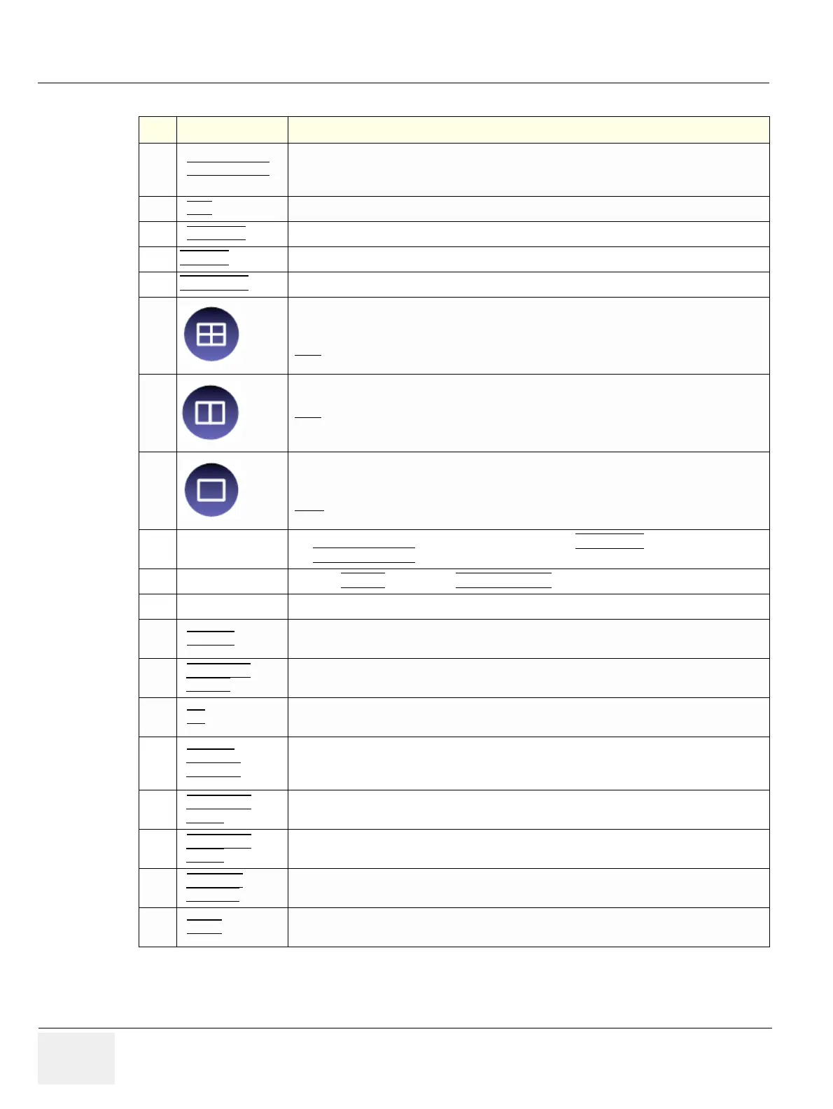
3
- Quarter size display of Sectional planes without 3D image or
- Quarter size display of Sectional planes + rendered 3D image
(Note:
The display depends on selected Acquisition- and Visualization Mode!)
4
Dual size display of Sectional Planes + rendered 3D image.
(Note:
The display depends on selected Acquisition- and Visualization Mode!
This format is not possible for Static 3D Acquisition)
5
- Full size display of a the reference image or
- Full size display of the rendered 3D image.
(Note:
The display depends on selected Acquisition- and Visualization Mode!)
6
Volume Box Position
and Volume Box Size
Adjust the Volume Box (ROI) Position resp. Size with the TRACKBALL
in the 2D Single image.
The upper trackball key
to change the Trackball function from Box Position to Box Size.
7
Start Acquisition Press the FREEZE
key resp. the right trackball key to start the Volume acquisition.
8
VISUALIZATION: After resp. during Volume Mode Acquisition:
• RENDER
After the 3D acquisition the system switches automatically to the read menu. The selected format
will be present on the monitor (e.g., 3D ROI Mode: sectional planes A, B, C + rendered 3D image).
• SECTIONAL
PLANES
After the 3D Sectional Planes acquisition the system switches automatically to the read menu.
The selected format will be present on the monitor (e.g., A,B,C - Sectional Plane mode).
• TUI
This method of visualization is consistent with the way other medical systems such as CT or MRI,
present the data to the user (slices through the data set, which are parallel to each other).
• VOLUME
ANALYSIS
The basic idea behind “VOCAL” is the combination of 3D ultrasound tissue (presented as voxels)
and the geometric information of surfaces in a 3D data set. After definition of contour in 3D space
(semi-automatically, manually or spherical) a wide range of functionality is given.
• SONOVCAD
HEART
Technology that automatically generates a number of views of the fetal heart.
At this time it can help to find the right and left outflow tract of the heart and the fetal stomach.
• SONOVCAD
LABOR
Allows to measure fetal progression during the second stage of labor – fetal head progression,
rotation and direction. Visual evidence and objective data of the labor process are provided.
• SONOAVC
GENERAL
This Feature can automatically detect low echogenic objects (e.g., follicles) in a volume of an
organ (e.g., ovary) and analyze their shape and volume.
• NICHE
Parts of the orthogonal sections A, B and C are compiled to a 3D-section aspect. The name
“Niche” has been chosen because the aspect shows a quasi spatial cut into the reference image.
Table 4-6 Pre-Volume Mode Functions
Step Task Expected Results