Analysis Protocols
Stratus OCT User Manual PN 2660021134133 A
6-24
Method of Optic Nerve Head Analysis
For each scan in the group, the Optic Nerve Head analysis detects the anterior surface of
the RNFL and the RPE. It detects the RNFL surface by searching each A-scan from anterior
to posterior until it finds reflectivity above a threshold value. From below the RNFL surface,
it searches each A-scan posteriorly for the highest rate of change in reflectivity to find the
RPE surface. Having determined these boundaries, the algorithm detects and measures all
features of disc anatomy based on the anatomical markers (disc reference points) on each
side of the disc where the RPE ends. It locates and measures the Disc Diameter by
tracing a straight line between the two disc reference points. It measures Cup Diameter
on a line parallel to the disc line and offset anteriorly by 150 micrometers (by default—Cup
Offset is adjustable). It determines Rim Area using the cup line as a posterior boundary;
for the rim lateral boundaries it uses lines extended from the disc reference points
perpendicular to the disc line and up to the anterior surface of the disc.
The results of these detection and measurement algorithms are displayed graphically on
th
e scan image. In the output display, you can adjust the placement of the disc reference
points, and thus the resulting measurements. Optic Nerve Head analysis then combines the
analysis and measurement of each individual scan into a composite image and
measurements of the whole optic nerve head.
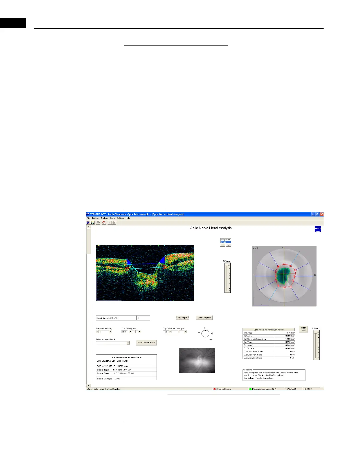
Output Display
Figure 6-15 Optic Nerve Head Analysis Output