Stratus OCT User Manual PN 2660021134133 A
Analysis Protocols
6-23
Output Display
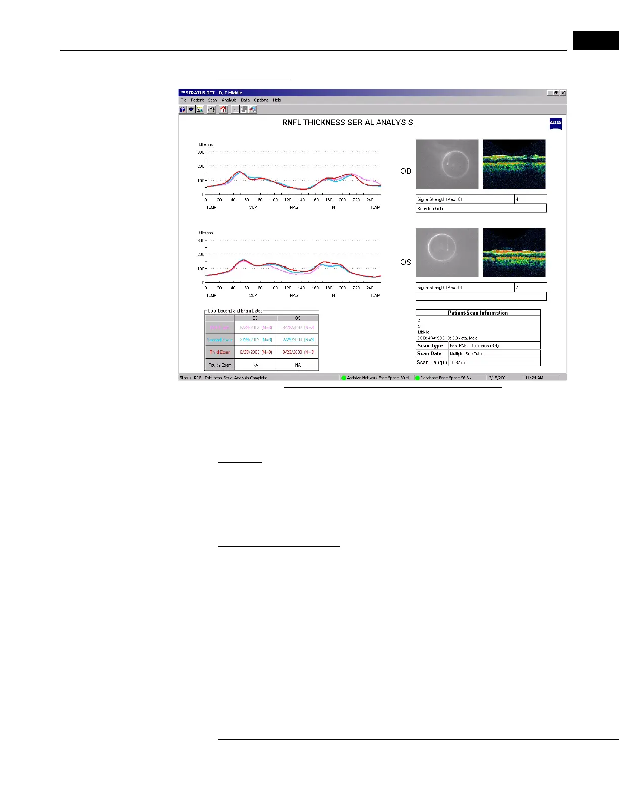
Figure 6-14 RNFL Thickness Serial Analysis Output
• The legend at bottom distinguishes the lines by exam date.
Optic Nerve Head
Application: Select Optic Nerve Head to access a multi-featured interactive analysis of the
optic nerve head. You can apply this analysis to one (F
AST) Optic Disc scan group at a
time. On the same display window, the output enables you to in
teractively assess and
measure the optic nerve—disc, cup, rim and cup/disc ratios—using each scan individually
and a composite of all scans.
Overview of the Analysis:
Using six radially acquired cross-sectional line scans, Optic Nerve Head (ONH) analysis
quantifies the amount of nerve fiber at the optic nerve head. It calculates two measures of
nerve fiber quantity. One is the cross-sectional area of the nerve fiber above the cup. This is
called the Rim Area. It is indicated in red on the individual radial scan (on the left side of
the screen). The other measure is the minimum distance between the RPE and the RNFL
surface. On each side of the nerve head, the analysis calculates this distance—indicated
as yellow lines—then averages them to yield the Average Nerve Width at Disc. The
analysis calculates these values for each of the radial line scans and then integrates them
to give results for the entire nerve head. The analysis screen presents the integrated values
on the right side along with a composite image of the nerve head constructed from all
scans.