Stratus OCT User Manual PN 2660021134133 A
Scan Acquisition Protocols
4-3
• X-Line
• Circle
• RNFL Thickness (3.4)—Glaucoma
• RNFL Thickness (2.27xdisc)—Glaucoma
• RNFL Map
• Fast RNFL Map—Glaucoma
• Proportional Circle
• Concentric 3 Rings
• Nerve Head Circle
• Optic Disc—Glaucoma
General Tips
The following information and usage tips apply to all scan protocols. Scan Protocol
Descriptions, Options And Tips, starting on page 4-6, provides a description for
each protocol, including its adjustment options and specific usage tips.
Scan Protocols Are Correlated with Analysis Protocols
When selecting a scan protocol, it is important to keep in mind the analysis protocol(s) that
you can apply to the resulting scan image. The analysis protocols are of two kinds:
Analyses and Reports (see page 6-13) and Image Processing Protocols (see page
6-37). Carl Zeiss Meditec designed the image processing protocols for use with any scan.
However, we designed each quantitative analysis protocol for use with a certain scan type
(line or circle) or scan pattern made on a certain retinal location (macula or disc). Several of
the scan protocols are designed for use with a limited subset of analysis protocols. An
analysis protocol might work on an unintended scan type, but even if it does, it may not
provide meaningful output. The table below shows the scan protocols for which we
designed each quantitative analysis protocol.
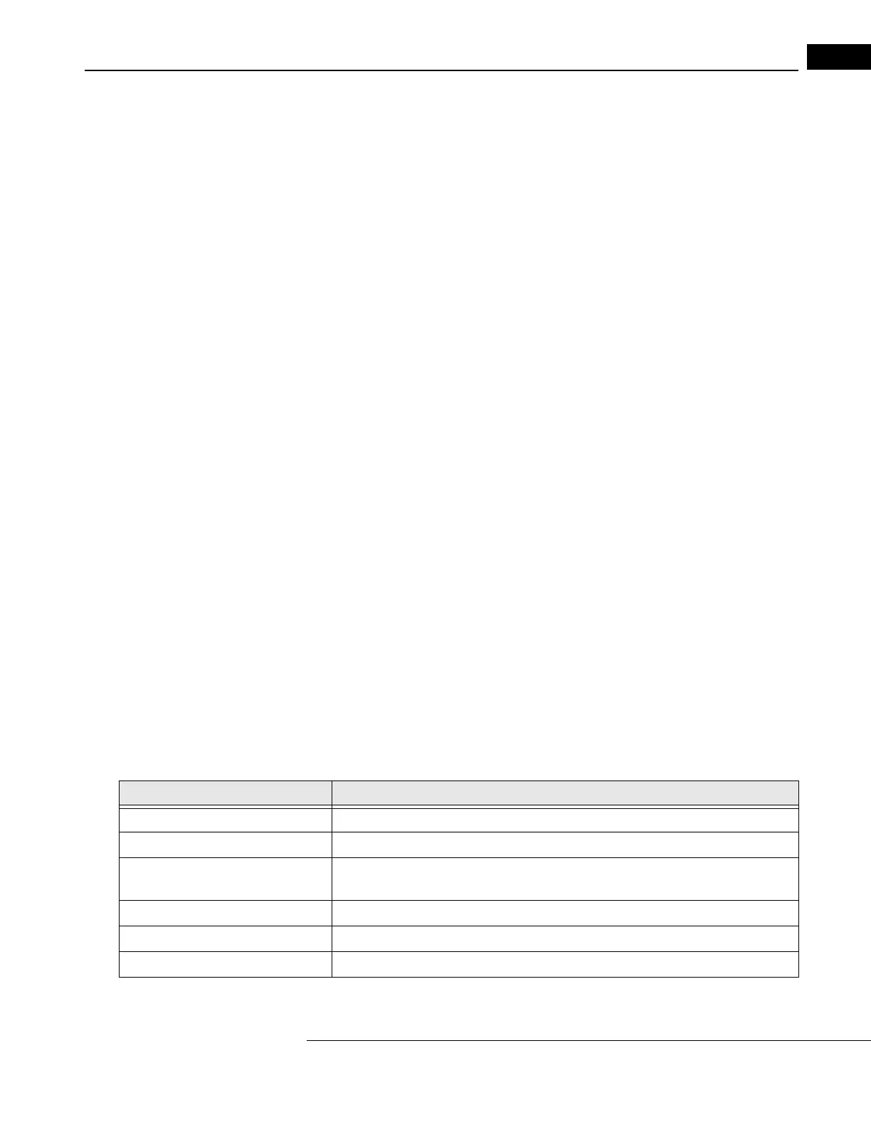
Table of Correlations
Analysis Protocol Designed for Scan Group(s)
Retinal Thickness Any scan protocol except (Fast) Optic Disc: 1 group on macula
Retinal Map Line, (Fast) Macular Thickness Map: 1 group on macula
Retinal Thickness/Volume Line, (Fast) Macular Thickness Map: 1 OS group and/or 1 OD group on
macula
Retinal Thickness/Volume Tabular Line, (Fast) Macular Thickness Map: 1 OS and/or 1 OD group on macula
Retinal Thickness/Volume Change Line, (Fast) Macular Thickness Map: 2 OS and/or 2 OD groups on macula
RNFL Thickness Any scan protocol except (Fast) Optic Disc: 1 group around disc