Scan Acquisition Protocols
Stratus OCT User Manual PN 2660021134133 A
4-8
repeated in several other protocols with variations in size and adjustability of parameters.
This protocol provides maximum adjustability, and you can use it to Define Custom
Scan
S (see page 3-16). The default pattern has 6 lines of 6 mm length. You can adjust the
length of all the scan lines by adjusting the aimi
ng circle size, or by adjusting the length of
the first scan in the series. After you save the first scan in the series, you cannot make
further adjustments.
Raster Lines
The Raster Lines protocol consists of a series of 6 to 24 equally spaced parallel line scans
over a rectangular region the size of which you determine. This general-purpose protocol
enables you to examine a rectangular region of interest on the retina more or less
thoroughly, depending on the size of the region and number of lines employed. The default
pattern has 6 lines over a 3 mm square. The scan series proceeds from superior to inferior;
each scan proceeds from nasal to temporal.
• You can adjust the height and width of the
aiming box, and the number of lines. The
height of the aiming box affects the spacing between the lines. The width of the
aiming box determines the line scan length. After you save the first scan in the series,
you cannot make further adjustments.
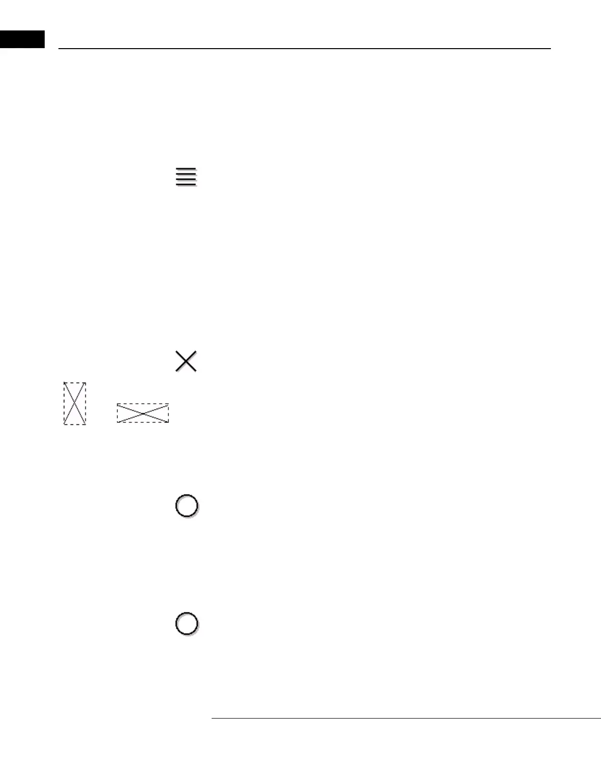
X-Line
Adjusting size of bounding box:
At left: increased height.
•The X-Line protocol consists of two line scans that intersect at their centers to form an
X. This general-purpose protocol is useful to examine a particular point of clinical
interest with two line scans that share a center point. You can average same-size
scans later and use this pattern to build custom scans. The default X pattern consists
of two perpendicular lines of length 3 mm. You can adjust line scan length by
adjusting the height and width of the imaginary box surrounding the X. Adjusting
either height or width affects the length and angle of both lines equally. After you
save the first scan, you cannot make further adjustments.
Circle
Select the Circle protocol to acquire multiple circle scans without returning to the MAIN
W
INDOW. Circle scans are normally applied around the optic disc (peripapillary region) to
measure nerve fiber layer thickness. This general-purpose protocol enables you to acquire
multiple circle scans, each of which you can repeat or tailor individually. You can average
same-size scans in later analysis. You can also use this scan to build a custom protocol. The
default pattern is a circle of radius 1.73 mm. You can adjust the radius of each scan.
RNFL Thickness (3.4)
The RNFL Thickness (3.4) protocol enables you to acquire three circle scans of diameter 3.4
mm around the optic disc. The only parameter you can alter is the number of A-scans—but
normative data applies only to scans acquired using the default 512 A-scans. The 3.4 mm
diameter circle represents a standard or typical size used to measure RNFL thickness. This