P1 Patient Monitor User’s Manual
10-3
brilirubin lamp, or sunlight;
Excessive patient movement;
Diagnostic test;
Low perfusion;
Electromagnetic interference, such as MRI device;
Electrosurgical equipment;
Concentration of nonfunctional hemoglobin, such as carbonyl hemoglobin
(COHb) and methemoglobin(MetHb);
The presence of certain dyes, such as methylene blue or indigo carmine;
Improper placement or incorrect use of pulse oximeter probe;
Shock, anemia, hypothermia or use of vasoconstrictor drugs, which can cause
blood flow in the arteries to drop to unmeasurable levels.
10.4. SpO
2
Display
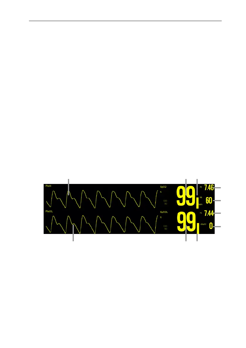
Wave form Display
(1) Pleth waveform (Pleth/PlethL): The amplitude of the Pleth/PlethL waveform can
directly reflect the strength of the patient’s pulse signal. The Pleth waveform is not
normalized.
(2) SpO
2
value (SpO
2
/ SpO
2
L): Percentage of oxygenated hemoglobin in relation to the
sum of oxyhemoglobin and deoxyhemoglobin.
(3) Pleth bar: Proportional to the intensity of the pulse.
(4) Perfusion index (PI): Gives the numerical value for the pulsatile portion of the
measured signal caused by arterial pulsation. PI indicates the signal strength of
1 23
4
5
6
7
3
21
 Loading...