Acclarix LX9 Series Diagnostic Ultrasound System User Manual
1. The eLV function is intended for use by the clinician who is qualified to use ultrasound for cardiac
function evaluations.
2. The measurement result is for reference only. The accuracy of the measurement result can be
affected by the image quality, the cardiac image section and procedures performed. The user is
responsible for the final decision.
3. Always confirm the detection of cardiac borders by visual inspection before storing the results in
the worksheet and report. If the result is in doubt, manual or other effective methods should be
applied to verify the correctness.
8.2.4.3.1 Auto EF
EF(Ejection Fraction) is evaluated by using two orthogonal apical planes: four-chamber (4CH) and
two-chamber (2CH) views, to provide automatic analysis of the global LV function.
Using Auto EF:
1. Scan the cardiac image in B mode, and acquire cine loops of 2CH or 4CH views.
Note:
It is required to acquire cine loops of at least 3 heartbeat cycles.
2. Freeze the ultrasound system.
3. Press the <Measure> key on the console to invoke the application measurement function.
4. Select Auto EF on the touch screen, and then select 4CH or 2CH depending on the cardiac cine
loops you acquired.
5. The ultrasound system starts to automatically analyze and calculate the EF results.
Note:
If the EF cannot be processed automatically because of improper image, the system will display
prompt message.
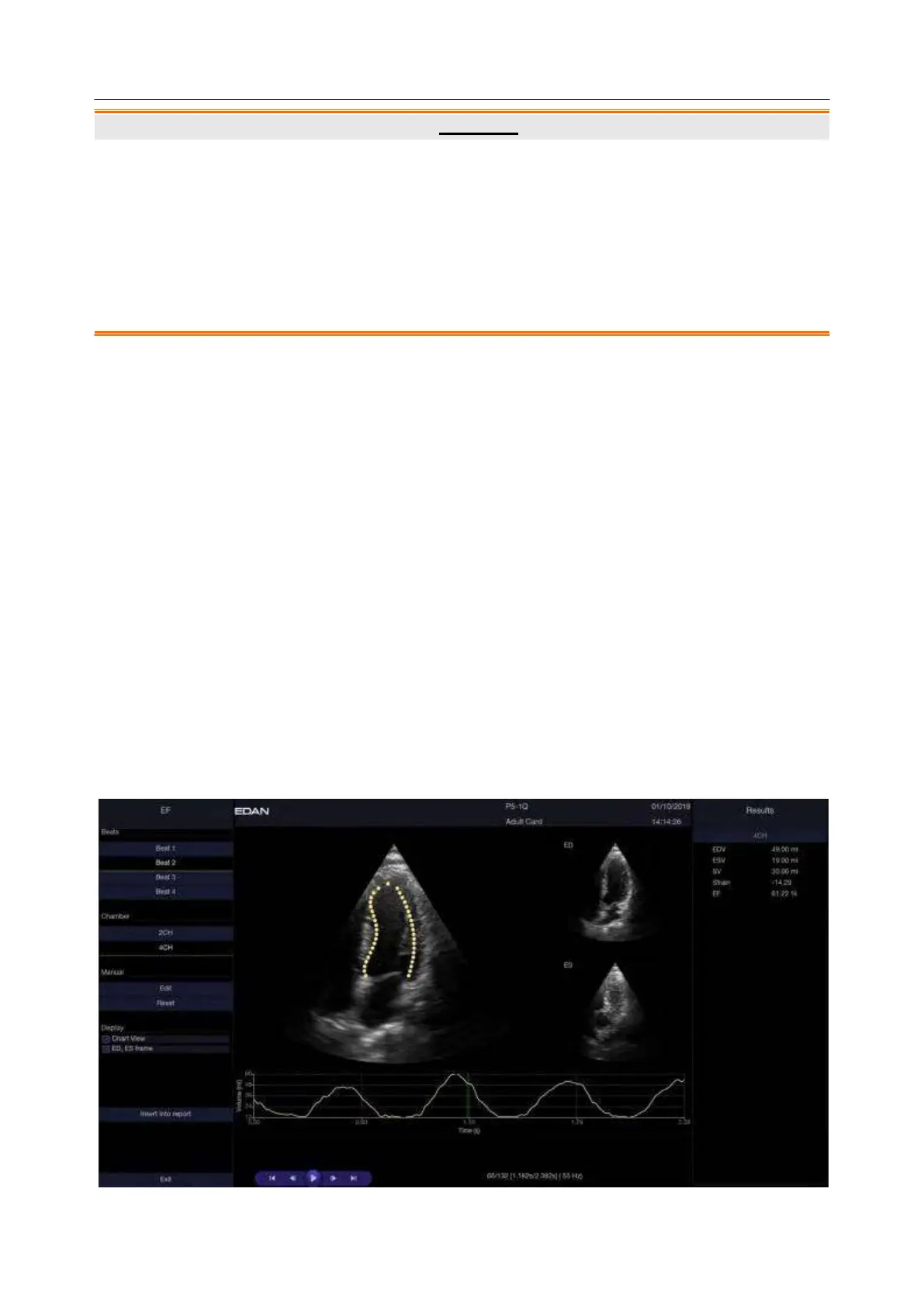
Understanding the EF screen:
 Loading...