TREATMENT STAGE
Manual Part Number
Update (Current)
Anatomy Strips
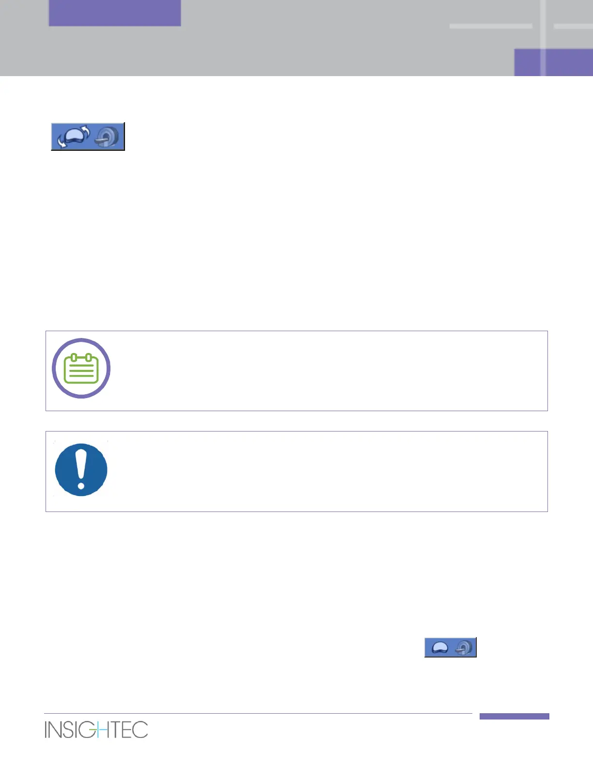
Click to acquire updated Current Anatomy strips (Axial and
Sagittal) and run the movement detection algorithm to compare
the Axial scans with the Axial Previous Anatomy scans (refer to
Section 7.2.8).
7.2.7. Spot Colors
The sonication spots are colored to provide additional feedback about their validity and status.
◼ Green – A valid spot that is ready to be sonicated.
◼ Yellow – Warns the user that the distance between the predicted dose of the spot and an LEDR is
below the recommended threshold.
◼ Red – The spot is invalid and cannot be sonicated. Either the spot is too close to an LEDR, or spot
parameters are not valid. Edit the spot to make it yellow or green.
When a spot is yellow or red, the reason will be displayed in the information box when
clicking and selecting the spot.
[N-25]
When a spot is Yellow, carefully review the spot and evaluate the risk versus the clinical
benefit prior to performing the sonication.
[C-6]
7.2.8. Current and Previous Anatomy Scans
While in Treatment stage, the Axial Current Anatomy scans effectively replace the original Axial Planning
images as the main working strip for updating the treatment plan (to account for prostate deformations –
see Section 7.2.10), planning new spots and evaluating the progress of the treatment.
After each sonication, the system automatically acquires a new set of Current Anatomy (both Axial and
Sagittal) images, which replaces the last acquired set (which is now renamed to Previous Anatomy).
To manually update the Current Anatomy strips (on-demand), press this button . This feature is
useful in case you suspect that movement (either of the probe and/or of the prostate) has occurred since
the previous update of the strips. Once the button is pressed, the system will: