EVALUATION STAGE
Manual Part Number
Click this button to prescribe and run an Axial Treatment
Evaluation scan, according to the range defined by the
Scanning Range lines. This scan will use the optimal central
frequency that was identified by the system.
When the scan ends, the new scanned MR series will appear in
the 3
rd
Exablate workstation image strip.
Click this button to select any series in the MR console browser.
This series is then uploaded to the workstation and can be
viewed in the 3
rd
image strip
Click this button to draw the area on all relevant MR images and
display its volume.
Show/Hide Volume
Measurement
Click on this button to Show/Hide the volume measurement
overlay.
8.3. Automatic Evaluation Scan Acquisition
1. Upon entering Evaluation Stage, the 3rd Image Strip is updated with the Sagittal Current Anatomy
strip, showing the Scan Range Lines as previously set in the Planning stage (see Section 6.5.2).
2. Either leave the Scan Range Lines as previously defined or edit them, as necessary.
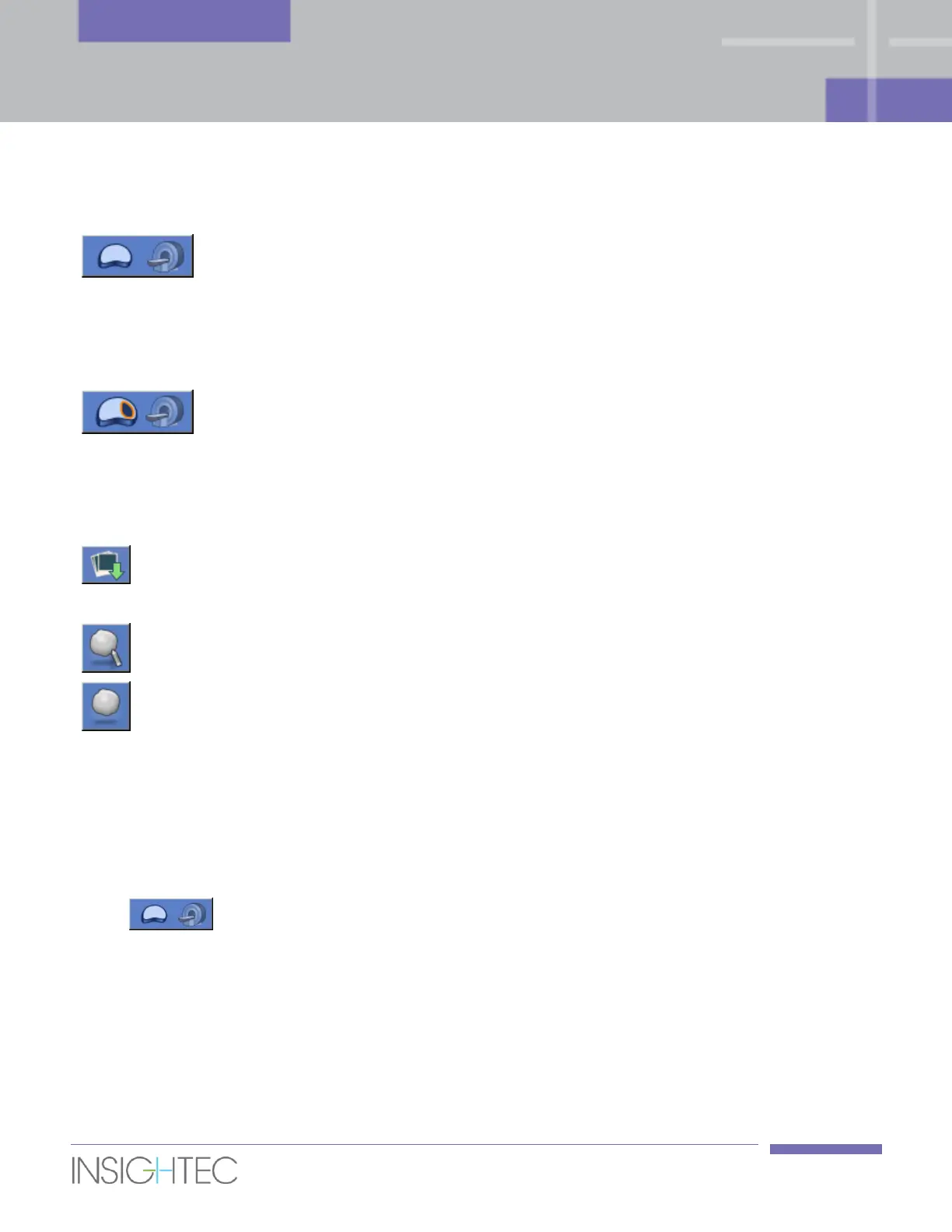
3. Click this tool to perform a T1w Axial Evaluation Scan (without contrast), covering the
range that is defined by the Scan Range Lines, and using the optimal Central Frequency value that
has been previously found (see Section 6.4.2).
4. The automatic Axial scan will be prescribed using recommended parameters (e.g., slice thickness of
3mm and zero spacing).
5. Once the MR completes scanning this series, the set of images will be retrieved automatically and
displayed in the 3rd Image Strip of the Exablate workstation.