In case the Planning images were acquired automatically through the Exablate
workstation (using Scan Prescribed Volume), the system automatically scans the Baseline
Anatomy series. Do NOT manually repeat the scan in such a case, as the Baseline Anatomy
strips will be replaced with the newly scanned ones.
[N-20]
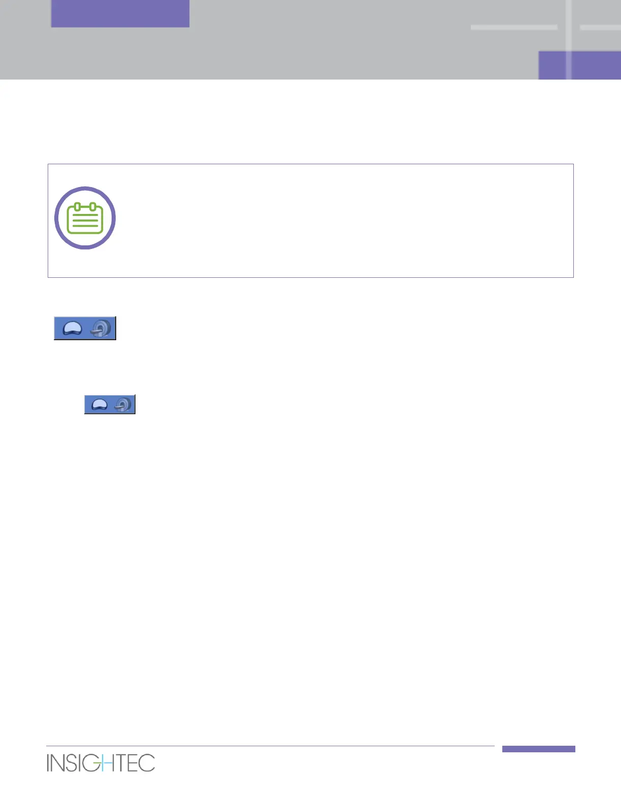
1. After manually loading Planning images, close the loading form.
2. Press this button to initiate the Baseline Anatomy scans.
3. The Axial Baseline Anatomy scan replicates the scan range and slice locations of the Axial Planning
images, while the Sagittal Baseline Anatomy scan includes 5-6 slices that are centered around the
treatment envelope.
4. Once the MR completes scanning the series, the set of images will be retrieved automatically and
displayed in the 2nd image strip (Sagittal Baseline Anatomy) and the 3rd image strip (Axial Baseline
Anatomy) of the Exablate workstation.
6.7. Defining a Treatment Plan
After loading the Planning images to the image strips and acquiring the Baseline Anatomy scans, you should
define the treatment plan by drawing the prostate capsule, rectal wall, region of treatment (ROT), and
detecting the balloon boundaries. Optional drawings include the target volume, and the following low
energy density regions (LEDRs): urethra, sphincter, nerve bundles and bladder.
Target Volume can only be drawn on a Diffusion-Weighted Imaging (DWI) series. All other manual
drawings are performed on the Planning images, while balloon boundaries detection is performed on the
Axial Baseline Anatomy images.