TREATMENT STAGE
Manual Part Number
Every sonication has a pre-set frequency direction, along one of the plane’s main axes. The frequency
direction is indicated by an orange arrow located in the lower right corner of the thermal image. In Multi-
Slice thermal imaging, the location of the thermal spot along the phase direction (which is perpendicular to
the frequency direction) is sensitive to imaging shifts which do not represent the true location of the
thermal spot.
The Exablate Prostate enables geometrical adjustments to be performed only in the S/I
direction. In case a significant shift is identified in the R/L direction, please contact your
service representative.
[N-36]
1. Select the Red Temperature Scaling Tool (Section 7.3.4) and reduce the temperature threshold until
a tight hot spot is clearly visible.
2. If a hot spot can be adequately identified, verify that it is within 1.0 mm of the planned location (the
spot circle). If it is, continue to the Thermal Dose Verification Procedure (see Section 7.4.4).
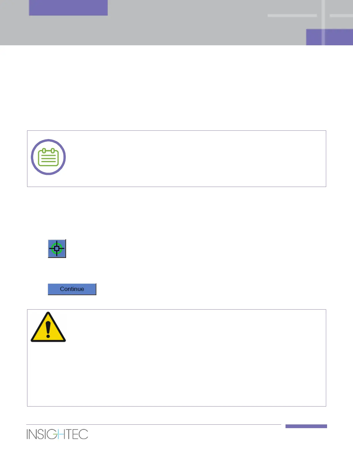
3. If the hot spot is over the 1.0 mm margin click this button, then click on the hot spot center
in the Selected Image window, to adjust to the correct position.
4. A pop-up message will show the required adjustment in spot’s location.
5. Click Accept or Reject adjustment and then click this button to return to the
Treatment Main Screen.
◼ Take extreme caution before performing an adjustment:
◼ If adjustment is required, it must be performed. However, do not perform an
adjustment unless you can clearly see the entire hot spot and be certain that the
adjustment is necessary.
◼ If the adjustment is over 2mm, before performing it apply another sonication with
a different frequency direction to confirm that the shift is real and not the result of
an MR central frequency shift.
◼ Failure to do so may increase the risk of treating unintentional tissue.