Stratus OCT User Manual PN 2660021134133 A
Acquire Scans
3-17
OCT Image Tab
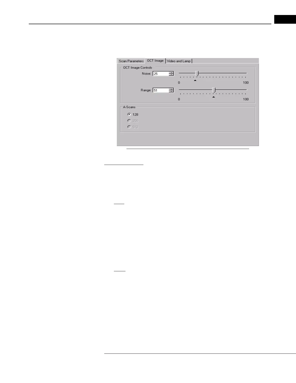
Click the OC
T Image Tab on the SCAN ACQUISITION WINDOW to adjust OCT image noise
and range values, or the number of A-scans for the scan protocol you have selected.
Figure 3-10 The OCT Image Tab (Scan Acquisition Window)
Noise and Range
These two sliders are set by default to filter the low end (background noise) and high end
(saturation signal) of the OCT interferometer signal. We recommend the default settings,
which are indicated by a mark on each slider, but you can adjust these settings to suit your
preference.
•
Noise refers to the level of signal that is considered background noise. At the default
setting, noise appears as random blue or green speckles in the black background. The
slider operates on a percentage scale: from 0, where nothing is considered noise and
so no signal is filtered out, to 100, where everything is considered noise and so all
signal is blocked. In effect, decreasing the noise adjustment increases the sensitivity
of the scanner: you get a stronger signal from the retina, but its clarity may be
compromised by noise. Conversely, when you increase the noise adjustment,
background noise decreases, but the intensity and definition of the scan image
decreases also. You must balance reduced noise against scan image quality.
•
Range refers to the range of interferometer signal levels depicted with the false color
scale in the Stratus OCT scan image. The upper end of the range sets the saturation
signal value. The scan image color scale depicts a range of relative signal values with
reference to the saturation value as the upper limit. When set properly, the saturation
value corresponds to the strongest reflected signal from the retina, and the color scale
brackets the entire range of the reflected signal strengths from the retina. If set too
high, the range of retinal reflectance is compressed into the cool colors of the scale,
or is off the scale below (depicted as black). This makes the retinal image weak or
nonexistent. If set too low, the range of retinal reflectance is compressed into the
warm colors of the scale, or is off the scale above (depicted as white, the saturation