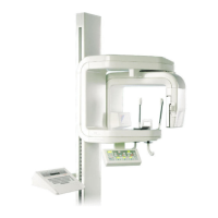
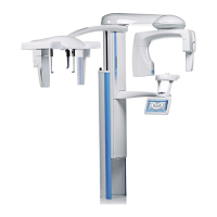
Chapter E - C-ARM AND IMAGING ARM
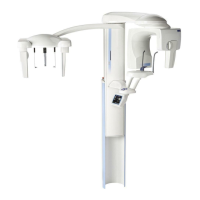
E-18 Planmeca ProMax
PANORAMIC BEAM AND PATIENT POSITIONING MECHANISM
ADJUSTMENT
Technical Manual
Panoramic beam vertical position
NOTE The lower border limiting plate should not move when the upper limiting plate is mov-
ing to its programmed position. Program the upper limit first.
If the X-ray beam is too low or too high, the vertical position of the beam must be
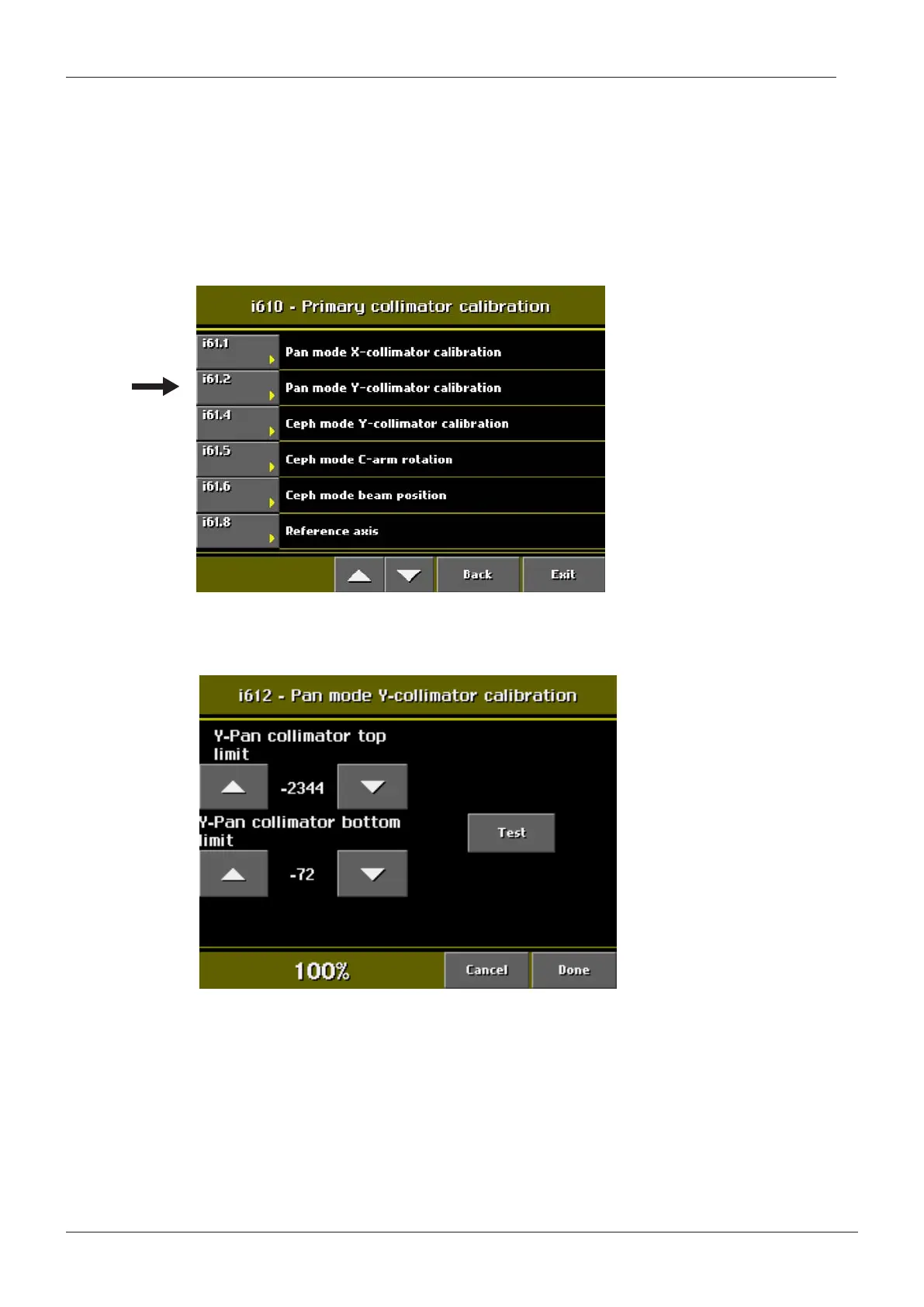
adjusted. From the display that appears when you selected Technical calibrations, select
Primary collimator calibration (i610). From the i610 display select Pan mode Y-collimator
calibration (i612).
Figure 24
The Pan mode Y-collimator calibration (i612) display appears.
Figure 25
Adjust the upper limit value of the X-ray beam with the Y-Pan collimator top limit arrow
fields. Drive the primary collimator to the selected position by pressing the Test field. Protect
yourself from radiation and press the exposure button to check the X-ray beam upper edge
position. If necessary, repeat the procedure.
Adjust the lower limit value of the X-ray beam with the Y-Pan collimator bottom limit arrow
fields. Drive the primary collimator to the selected position by pressing the Test field. Protect
yourself from radiation and press the exposure button to check the X-ray beam lower edge
position. If necessary, repeat the procedure. Accept the new position and exit the calibration
mode by touching the Done field.
 Loading...