Chapter E - C-ARM AND IMAGING ARM
Planmeca ProMax E-39
PANORAMIC BEAM AND PATIENT POSITIONING
MECHANISM ADJUSTMENT
Technical Manual
2.9 Taking a ball phantom exposure
NOTE Attach the ball phantom to the adapter on the patient support table.
NOTE If the temple supports are in place, remove them before taking a ball phantom exposure.
Take a ball phantom exposure to check that the X-ray beam, the patient support table and the
C-arm are correctly adjusted.
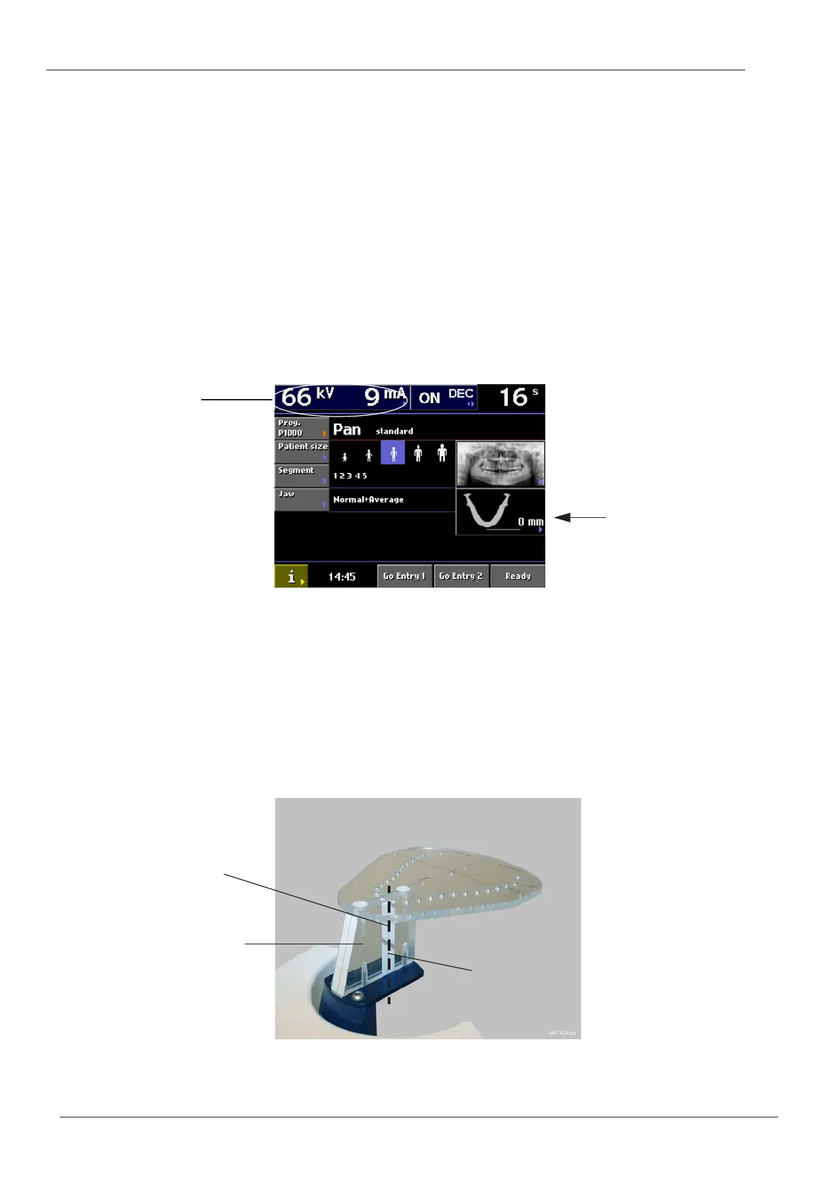
On the X-ray unit main display, select following settings:
- standard panoramic program
- full exposure area (no segmentation)
- small adult (third patient size symbol)
Selecting these settings will automatically change the exposure values to 66 kV and 9 mA.
Manually set the exposure values to 60 kV and 2mA.
Figure 52
Using the thumb wheel on the underside of the patient support table, position the layer light
so that the light beam hits the reference line on the ball phantom support plate. The three
patient positioning lights will be automatically switched on.
NOTE The layer position value on the X-ray unit main display must be zero (0) as shown above
when the layer light beam is positioned on the reference line . If this is not the case, you
will have to calibrate the layer light on display i660. Refer to section 2.10 “Panoramic
mode patient positioning lights” on page E-41 for details.
Figure 53
Set exposure values to
60 kV / 2 mA
Layer position value
Ball phantom
support plate
Reference line
Layer light
beam
 Loading...