USER MANUAL (Instruction for Use) SW 6.1
Device / System Description
Version 6.1.6 dated 2020-01-31 EN Page 50 of 201
movements. The lateral x-y- movements seen by the eye tracking system are calculated, with the
help of an eye model, into eye rolling. The scanner of the system is moved according to this
Rotation Balance with the Eye Tracker.
Changes of the pupil center during the treatment are corrected using additional limbus tracking
and pupil center shift control (PCSC) with both SCHWIND AMARIS models.
As an optional feature for all SCHWIND AMARIS models, the tracker can compensate for
cyclotorsional eye movements (5
th
dimension), dynamically during the ablation process and
statically with reference images taken during diagnosis with corneal or ocular wavefront
analyzers. The resulting angle is automatically calculated and compensated in the ablation
profile.
As an additional option for the SCHWIND AMARIS 750S and 1050RS models Z- movements (6
th
dimension) can be measured and actively compensated during the ablation process. Moreover
AMARIS systems equipped with a 6D- eye tracker will actively measure eye rolling movements
which can be visualized in the treatment screen.
Additionally, the model AMARIS 1050RS offers the option of a Latency Free Eye tracking feature
as 7
th
dimension.
The eye tracker works with IR-light illumination, thus making it independently from any density
setting of the OP-field illumination or room illumination.
As an additional option, an online pachymeter (OCP) can be integrated for measuring the central
corneal thickness before, during and after the treatment. The OCP control and data collection is
integrated in the laser software, which also ensures the matching of data for each treatment.
The user control of the machine is based on the WindowsXP
TM
professional operating system,
installed on a Panel-PC with touch screen for sterile use through the application of re-sterilisable
pens. The PC can be adjusted to a suitable working position for both surgeon and assistant.
The planning and calculation of any treatment is based on the SCHWIND eye-tech-solutions CAM
software (Custom Ablation Manager) as an independent software module. Treatments can also
be planned at the SCHWIND diagnosis workstations of the corneal or ocular wavefront analyzer.
The control of the system is based on several local control units and a main control unit, which
is a DebianLinux operated embedded PC.
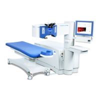
An operation microscope, with which the surgeon can observe the patients eye during the whole
treatment process, is integrated in the laser arm. The microscope is designed with an optimized
stereopsis angle of 14° and equipped with a 5x motorized magnification changer, which can be
changed manually or through software control. An external camera can be mounted on the
microscope for video observation by additional personnel or for video documentation. For future
purposes the microscope is prepared for coupling-in of alpha-numerical or graphical information
into one of the eyepieces.
Four high power white-light LEDs illuminate the OP field for best visual inspection of the patient's
eye with the microscope. The illumination density can be set manually by the user or through
software control.
For additional check of the patient's eye, e.g. control of the flap repositioning, an optional slit
lamp can be mounted on the laser arm.
At the left and the right sides of the microscope are key pads for easy access to the most
commonly needed manual settings, such as illumination density, microscope magnification and
particle aspiration. Necessary confirmations can be made here or through pressing “OK”
 Loading...