USER MANUAL (Instruction for Use) SW 6.1
Device / System Description
Version 6.1.6 dated 2020-01-31 EN Page 56 of 201
4.9 Microscope and Illumination
A coaxial stereo microscope is used for exact control and focusing of the corneal surface. It allows
coaxial stereoscopic inspection of the eye without the control beam path being guided through
the working optic of the device. Therefore, a very high quality picture is possible.
Abrasion of the epithelium and LASIK can be performed under optimal control conditions.
Furthermore, the inspection of treatment results is possible.
The crosshair is aligned and dimensioned for the magnification changer in position 1.0.
Any another magnification could result in a shift of the crosshair. Further function description of
the operation microscope is provided in chapter 6.8 Operating of the Microscope.
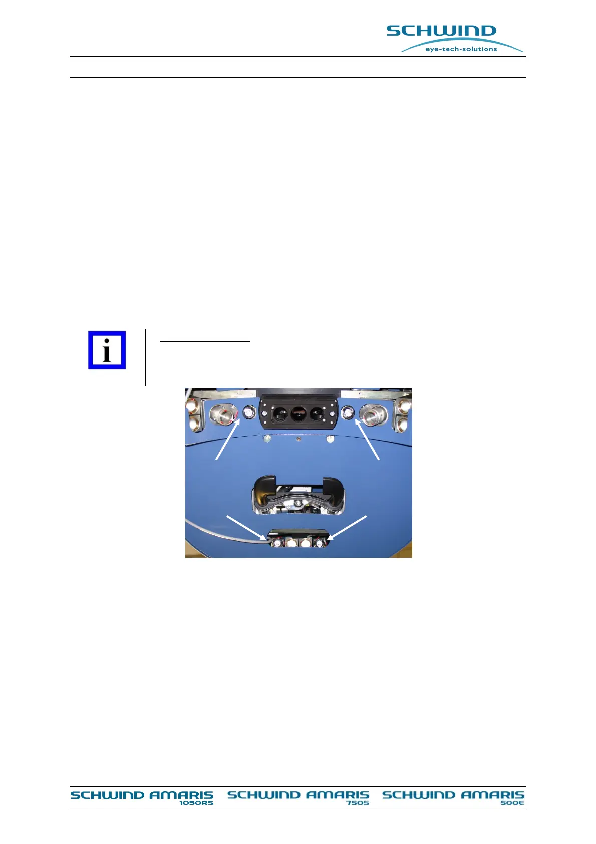
For illumination of the treatment area, a LED illumination system (see Figure 4-3: LED
illumination) is integrated into the unit. This white light illumination can be adjusted in brightness
and switched off and on with the illumination control on the AMARIS control panel, if desired.
Brightness can be adjusted for best treatment observation combined with short application time.
IMPORTANT NOTE
High brightness can be very unpleasant to the patient, so avoid inappropriate
light illumination and reduce time of illumination to treatment time.
Figure 4-3: LED illumination
4.9.1 Major Components
The major components of the operating microscope are:
Motor-driven 5 step magnification changer
10° to 50° microscope binocular tube (1)
4.9.2 Magnification Changer 5 Step
The 5 step magnification changer (5) is motor-driven. The magnification can be changed step-
by-step manually by using the appropriate buttons <+> and <—> at the key pad (5), or
automatically controlled by the software (see Figure 4-4: Components of the operation
microscope (example AMARIS 750S).