Iris Diagnostics, a Division of Iris International, Inc.
iQ
®
200 Sprint™ (2G) Automated Urine Microscopy Analyzer Service Manual 300-4949 Rev A 01/2005 3-16
3. Components
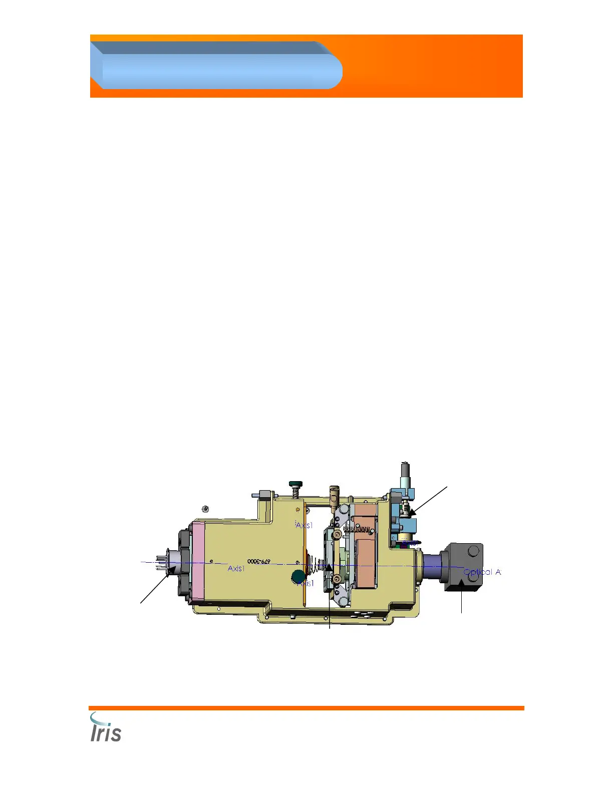
Optical Bench Assembly #700-3500
Sample is presented as a lamina sandwiched between enveloping layers
of suspending fluid. The fluids pass into the Flow Cell in front of a
microscope. The lamination positions the specimen exactly within the
depth of focus and field of view of the objective lens of the microscope. A
CCD (charge coupling device) video camera, connected to the
microscope, captures five hundred frames per sample, as each
microscopic field of view is illuminated by the flash of a strobe lamp. The
resulting pictures are digitized and delivered to the Analysis Processor
computer.
The Optical Bench Assembly (OBA) is a mechanical/electro-optical
assembly that provides for the microscopic images of a transmissively-
illuminated ultra-thin sheet of the specimen to be captured as a video
image by the CCD camera. Three thermistors are mounted on different
component parts of the OBA to check that the temperature is stable.
When temperatures are changing, because of the sub-micron accuracy
required for proper images the OBA requires re-focusing.
The mechanical part of the OBA includes:
1. Flow Cell
2. Strobe Light
3. CCD camera
4. 20 x Objective
5. Auto Focus Motor
CCD camera
Auto Focus Motor
Flow Cell
Strobe Light
 Loading...