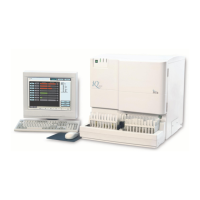
Iris Diagnostics, a Division of Iris International, Inc.
iQ
®
200 Sprint™ (2G) Automated Urine Microscopy Analyzer Service Manual 300-4949 Rev A 01/2005 3-17
3. Components
Flow Cell # 525-3077
This precision assembly is where the thin sheet of liquid specimen is
presented in a continuous flow microscope-slide manner for observation
by the image capturing system. The sample is pulled into the Flow Cell
by the Evacuation pump. Lamina encases the sample, keeping the
specimen in thin sheet, and keeps the specimen away from the Flow Cell
surfaces to minimize surface contamination.
Strobe Light # 065-0059
The strobe light provides short, high-intensity, parallel-light strobe-light
pulses on a controlled basis through the thin sheet of specimen. This
subsystem includes a 400V to 1000V strobe power supply and a high
intensity strobe flash tube.
CCD Camera # 700-3565
The CCD camera, coupled to the Microscope, takes magnified high-
resolution digital pictures of the sample going through the Flow Cell.
20 x Objective # 220-3009
The objective magnifies the sample going through the Flow Cell.
Autofocus Motor ” Micro-nudger” # 700-3505
This mechanism allows moving the Flow Cell into the appropriate position
where the thin specimen is focused for image capture. Focus position is
achieved with an ultra-fine-pitch stepper motor providing a movement of
about 0.2 microns per full step.

 Loading...