Iris Diagnostics, a Division of International Remote Imaging Systems, Inc.
iQ
®
200 Sprint™ Automated Urine Microscopy Analyzer Service Manual 300-4949 Rev A 01/2005 4-26
4. Adjustments
Flow Cell Lateral Adjustment
1. Place one glass tube in position 1 of a routine rack. Fill the tube with
3 mL of positive control. Do not apply barcode labels on the tube.
2. Place the rack on the right side of the sampler and press GO.
3. Press the Windows key on the keyboard.
4. Select Program – Accessories - Window Explorer.
5. Select C: - Iris2k1 – Bin. Double-click on Runfile Analyzer file.
6. Select the following options:
• Show Part in Fr (particles in frame)
• Focus Est (focus estimator)
• Frame - ALL
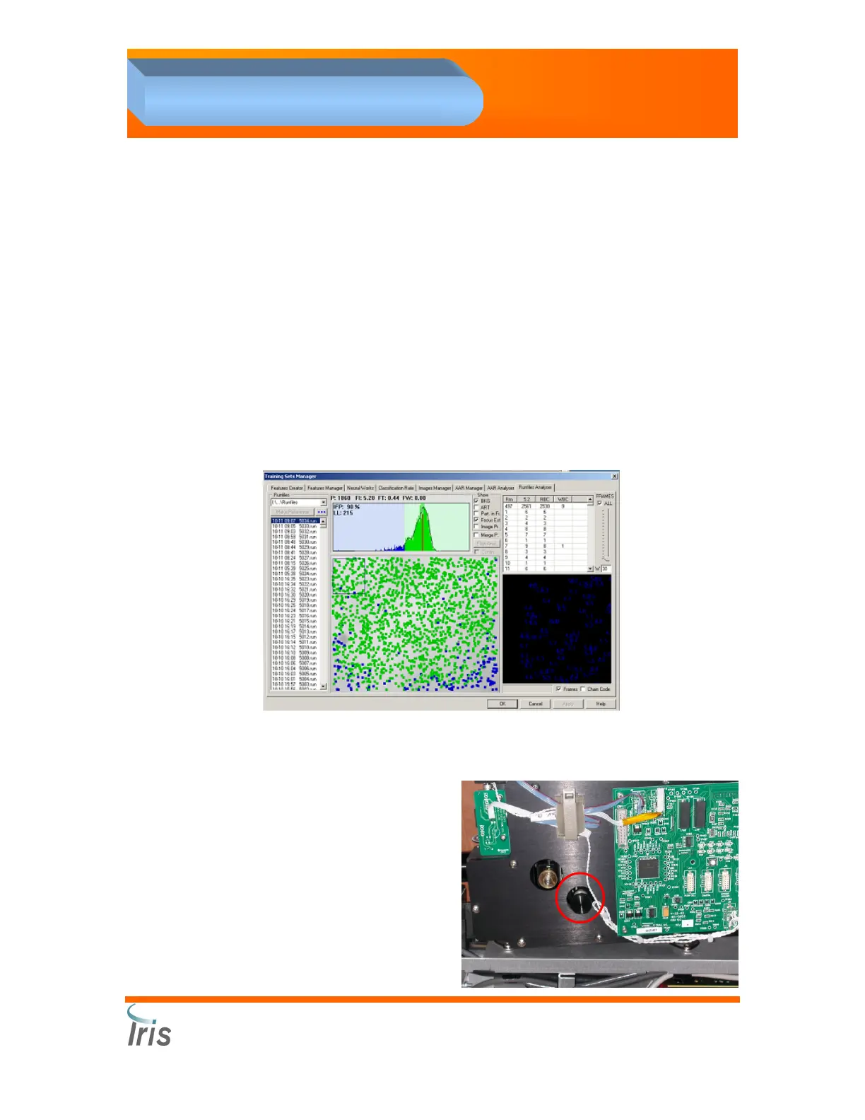
7. Preview the runfile to evaluate the cell distribution on the screen.
8. Below is an example of the image you should see of a correct lateral
adjustment.
NOTE: If the flow cell is not properly aligned, you will see an area within
the field that will NOT display any cells.
9. If a lateral alignment is
needed, use the knob
highlighted on the right.
 Loading...