Chapter H - CEPHALOSTAT
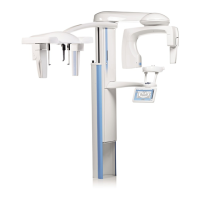
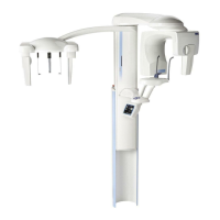
Planmeca ProMax H-47
ADJUSTMENTS AND CALIBRATIONS
Technical manual
Calibrating the sensor head, automatic procedure
NOTE Use the high speed feature.
Protect yourself from radiation and press the exposure button.
Select Ceph All Binnings from the Calibrate menu. Do not release the exposure button.
Figure 83
Press and hold down the exposure button for the duration of the exposure cycle. After the
exposure cycle the image from the latest exposure (3x3; enhanced resolution) is shown in the
Dimax2Tool window. The triangle on the image must be even, without any irregularities. If it is
not, edit the triangle as described on page page H-45. The calibration must be performed with
4x4 (normal resolution) binning manually as described in section “Calibrating the sensor
head, manual procedure” on page H-43.
Editing the calibration image
In case the calibration image contains horizontal stripes, the image can be edited, i.e. the
selected row can be removed by marking it as “bad”. You can enlarge the image with the
zoom function if needed. The image can be moved with the Hand-tool.
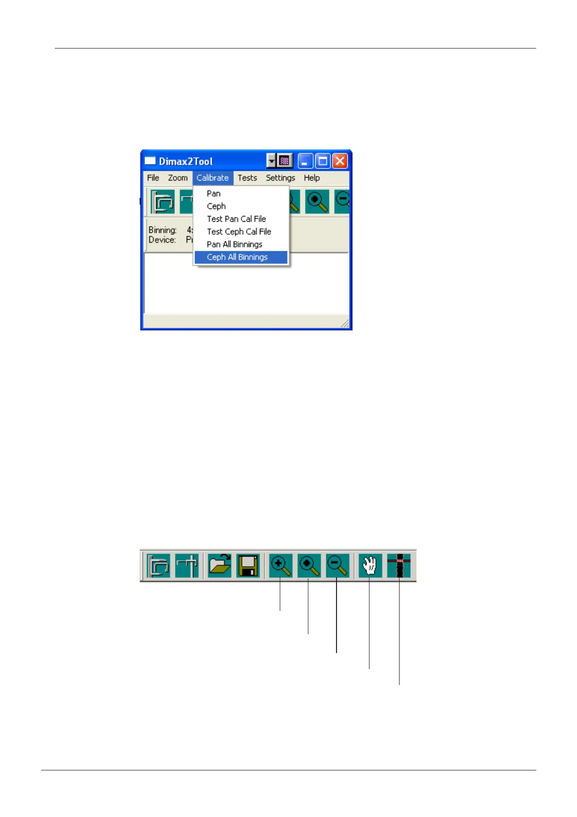
Press the Marking tool button located on the right side of the toolbar and then click the row
that you want to mark with the left mouse button. Note, that the row number is shown on the
status bar at the bottom of the window.
Figure 84
Zoom in
Fit image to window
Zoom out
Hand
Marking tool
 Loading...