6 Acquiring Images
Basic User Manual 71
1. Optimize a B-mode image.
2. Press the
CFM
key on the control panel, and then tap
PDI
on the touch screen to enter the PDI mode.
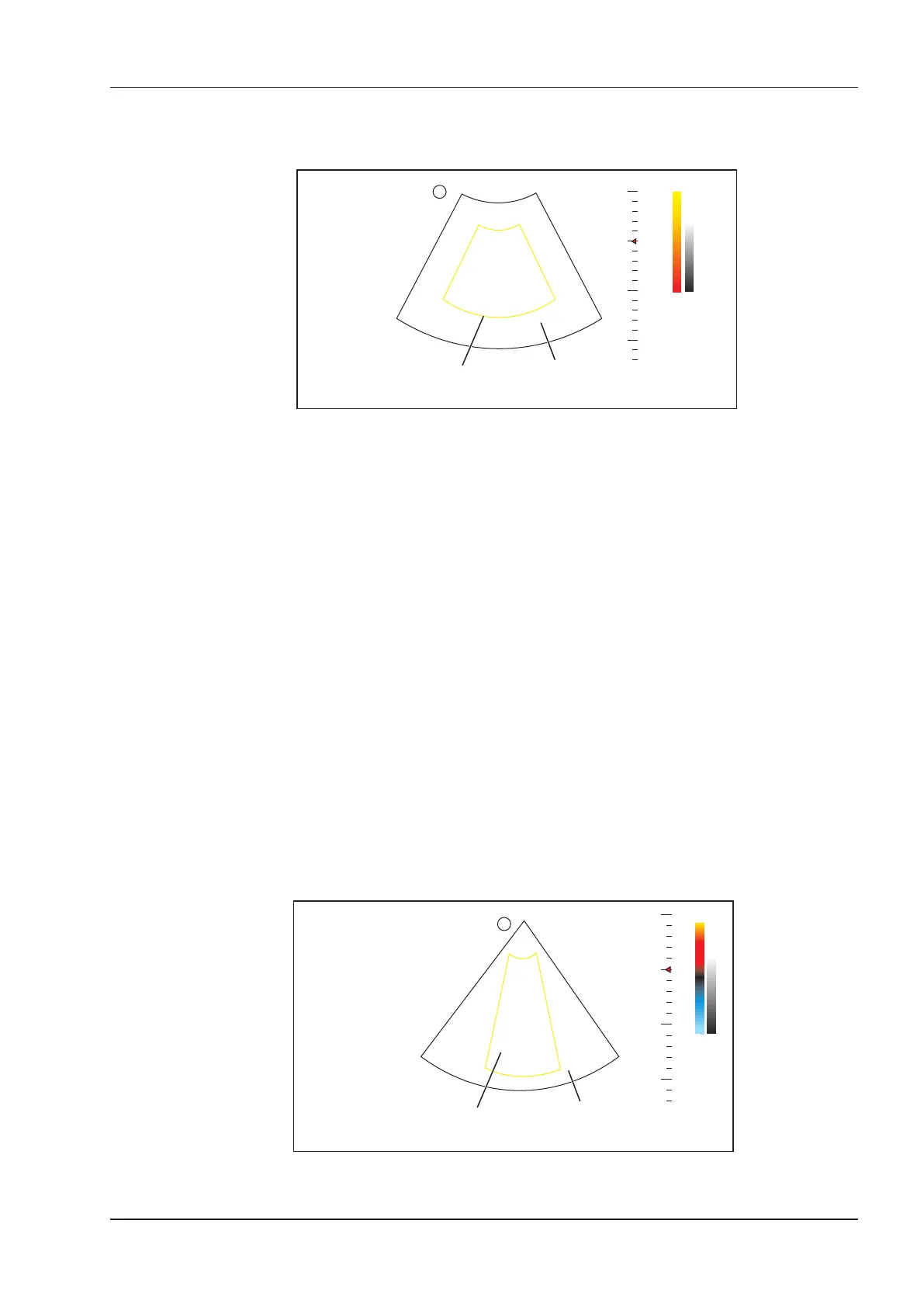
0
5
10
15
2D Imaging
Color Flow ROI
B
FPS 47
D/G 3/1
GN 255
I/P 3/30
PWR 70
FRQ 3-4.8
D 16.5cm
C
PRF 1.0
WF 86
GN 255
C/P 1/50
PWR 70
FRQ 2.2
S
Figure 6-7 PDI-Mode Imaging Screen
3. Adjust color ow ROI.
−
Move the trackball to position color ow ROI.
−
Press the conrm key on the control panel to adjust the size of color ow ROI.
−
Press the conrm key again to reposition color ow ROI.
4. Optimize the PDI-mode image. For details, refer to Section 6.3.4 Optimizing CFM/PDI/TDI Mode Images.
5. Tap
PDI
again to exit the screen.
6.3.3 TDI Mode
NOTE:
TDI imaging is only applied to cardiac applications by using the phased array probes.
TDI (Tissue Doppler Imaging) is a color ow imaging technique which detects the low frequency signal reected
from the cardiac muscle. TDI provides the ow information of velocity and direction for cardiac movement.
TDI uses the low velocity and the high amplitude adjusted from the wall filter to create a color-coded tissue
imaging.
Perform the following steps to acquire TDI-mode images.
1. Optimize a B-mode image.
2. Press the
CFM
key on the control panel, and then tap
TDI
on the touch screen to enter the TDI mode.
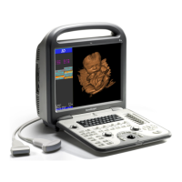
0
5
10
15
22
-22
cm/s
2D Imaging
Color Flow ROI
B
FPS 47
D/G 3/1
GN 255
I/P 3/30
PWR 70
FRQ 3-4.8
D 16.5cm
C
PRF 1.0
WF 86
GN 255
C/P 1/50
PWR 70
FRQ 2.2
S
Figure 6-8 TDI-Mode Imaging Screen