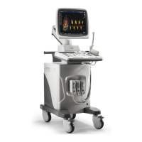
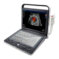
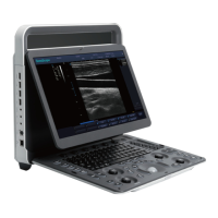
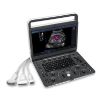
6 Acquiring Images
78 Basic User Manual
Tap the left or right part of
M Process
to adjust the setting.
■ Sweep Speed
Sweep speed is used to set the sweep speed of the M trace. A faster speed is more suitable to view the motion.
To adjust the sweep speed:
●
Tap the left part of
Speed
to decrease the value.
●
Tap the right part of
Speed
to increase the value.
■ Chroma
Chroma is used to colorize the gray scale image to enhance the discrimination capability.
To adjust the chroma:
Tap the left or right part of
Chroma
on the touch screen to adjust the setting.
■ Display Format
Display format is used to view the image better.
To set the display format:
Tap the left or right part of
Display
on the touch screen to adjust the setting.
■ Power
Power is used to select the amount of ultrasound acoustic power produced by the probe. The adjustment range of
the power is 30%-100%, and ±10% can be adjusted each time. The real time value of the power is displayed in the
imaging information area on the basic screen.
To adjust the power:
●
Tap the left part of
Power%
on the touch screen to decrease the value.
●
Tap the right part of
Power%
on the touch screen to increase the value.
NOTE:
Expose the patient to the lowest practical transmit power level for the shortest possible time to achieve a
satisfactory diagnosis.
■ Video Invert
Video invert is used to invert the M trace display related to brightness.
To enable or disable the video invert:
●
Tap
Video Invert
on the touch screen to enable the feature.
●
Or, tap
Video Invert
again on the touch screen to disable the feature.
6.5 Acquiring Spectral Doppler Images
Spectral Doppler imaging is intended to provide measurement data concerning the velocity, the direction, and the
category of the arterial or vein ow. It contributes a more accurate qualitative analysis than the color ow imaging.
Spectral Doppler includes Pulsed Wave Doppler (PW) mode and Continuous Wave Doppler (CW) mode.
6.5.1 PW Mode
Pulsed Wave Doppler (PW) is a Doppler mode that measures velocity in a PW sample volume and displays
that information in a spectral trace with audio output. PW includes conventional PW and High Pulse Repetition
Frequency (HPRF). PW Doppler can be combined with the B mode for rapidly selecting the anatomical site for PW
Doppler examination. The site where PW Doppler data is derived appears graphically on the B-Mode image.