6 Acquiring Images
76 Basic User Manual
0
5
0
10
10
5
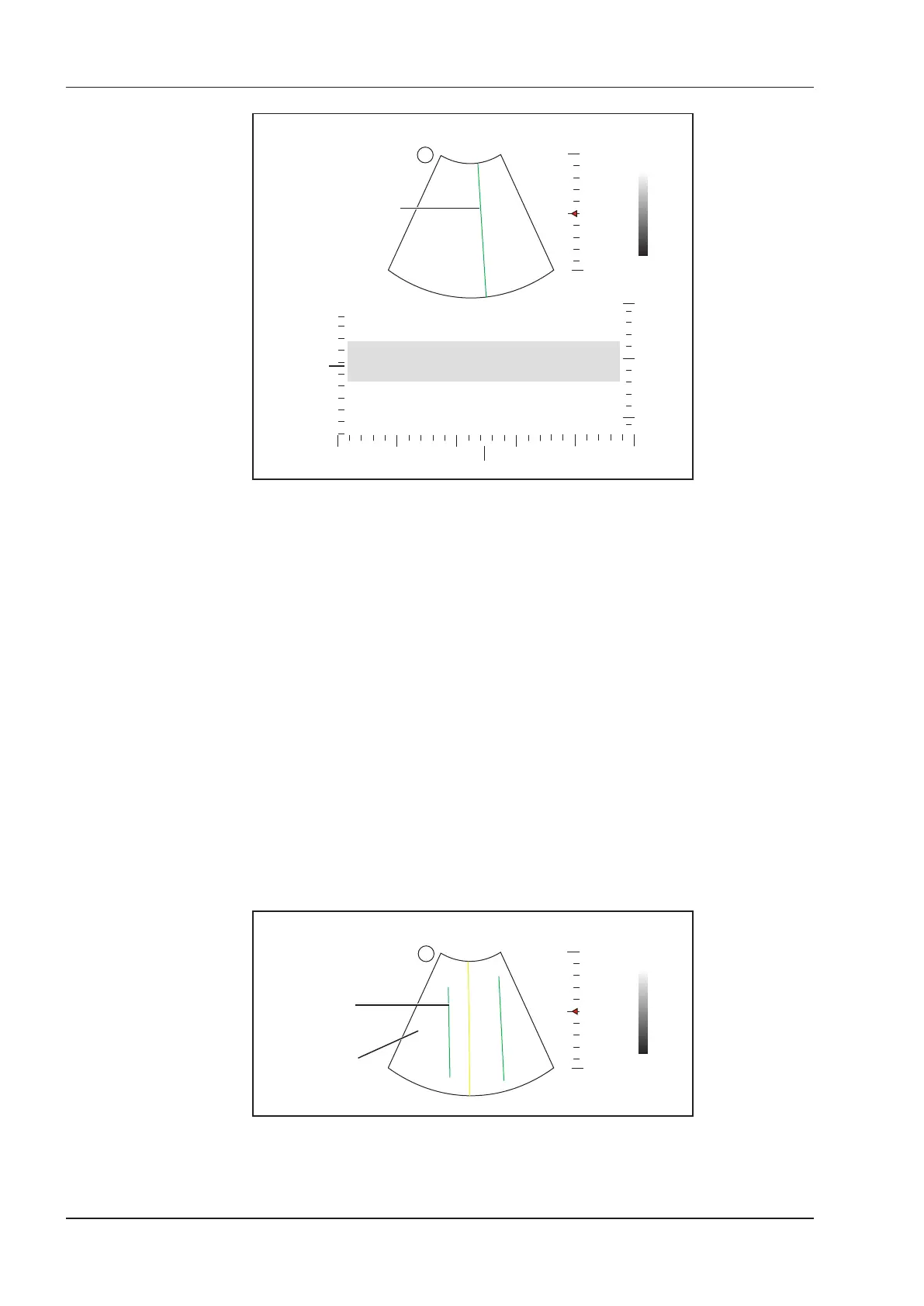
Y-axis: depth
X-axis: time
S
M-line
Figure 6-11 M-Mode Imaging Screen
−
X-axis is the time scale.
−
Y-axis is the depth scale.
NOTE:
Once the M mode is activated, you can move the trackball to stop the M trace and adjust the M line. The
system continues tracing if the trackball is idle for more than 0.5s.
5. Optimize M-mode image. For details, refer to Section 6.4.3 Optimizing M-Mode Images.
6. Press the
M
key again to exit the screen.
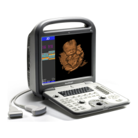
6.4.2 Anatomical M-Mode
Anatomical M-mode is used for fetal cardiac applications. Anatomical M-mode can be used with phased array
probes when performing cardiac exams or convex probes when performing abdominal exams. In the anatomical
M-mode, the M-mode cursor can be positioned perpendicular to the anatomical structure and be adjusted 360° even
when viewing motion patterns for irregular objects. It is used to study the ventricular function of the heart.
Perform the following steps to acquire anatomical M-mode images.
1. Enter the inactivated B+M mode, tap
Steer M
on the touch screen to set the number of M lines.
Multiple M-lines display after selecting the desired number as the following gure shows.
0
5
10
M-line
2D Imaging
S
Figure 6-12 Inactivated Anatomical M-Mode Imaging Screen
2. Adjust the position and the angle of M line.
−
Press the conrm key on the control panel to select the desired M line.