F15 Series Fetal & Maternal Monitor User Manual Fetal Monitoring
- 92 -
WARNING
1 Before insertion of IUPC, placental position should be confirmed, amniotic membranes are
adequately ruptured and sufficient cervical dilatation is assured.
2 Try to insert the catheter opposite the placental site. Do not insert the introducer beyond the
cervical OS. Use it with caution when uterine infection is present.
3 If resistance is met at any time during insertion, withdraw the catheter slightly and try at a
different angle. Forced insertion may result in patient’s discomfort or injury.
4 Do not catheterize if placenta previa is diagnosed, or if uterine bleeding from an
undetermined source is present.
CAUTION
1 Since procedures vary according to hospital needs/preferences, it is the responsibility of
the hospital staff to determine exact policies and procedures for both monitoring and
amnioinfusion. The safe and effective use of the IUPC depends on the skill of the clinician
who applies /uses it.
2 The IUPC has been sterilized by gamma radiation and is sterilized and non-pyrogenic
unless package is broken or open. Do not re-sterilize it.
NOTE:
Refer to the instruction on the package for more information about using the IUPC.
7.7.3 IUP Monitoring Procedure
1) Insert IUPC using the procedure described in section 7.7.2 Directions for Use of IUPC.
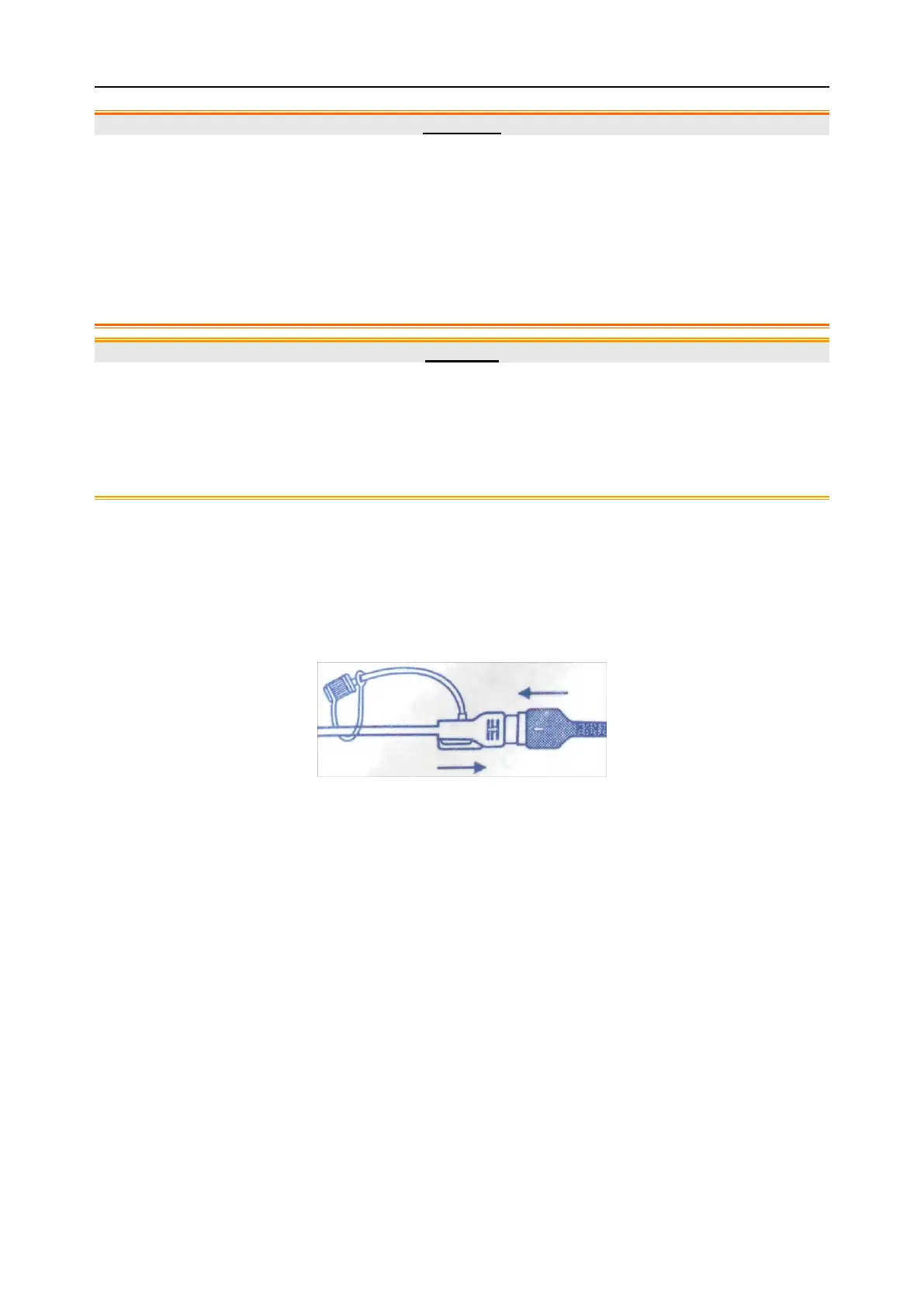
2) Connect the IUPC to the DECG-IUP integrated cable.
Figure 7-12 Connect Catheter to DECG-IUP Integrated Cable
3) Connect the DECG-IUP integrated cable to the DECG Fetal&Maternal Module.
4) Connect the DECG Fetal&Maternal Module to the fetal monitoring socket of the monitor.
5) Ask the mother to cough. A spike on the trace in response to the cough indicates proper
positioning and function of the IUPC.
6) Wash timely during monitoring. A spike on the tracing will respond to the washing.
7.8 Monitoring Fetal Movement
7.8.1 Auto Fetal Movement (AFM) Monitoring
During fetal heart monitoring with ultrasound, the fetal movement signals are also detected. The fetal
movement signals differ from the Doppler heart rate signals in that they have larger extent and lower
frequency. The larger extent is because of the bigger scope of moving areas (e.g., the fetal arms or
legs); lower frequency is because of the lower velocity of the fetal movements compared with those of
the fetal heart.
Only US1 channel can perform AFM. But be aware that when monitoring twins or triplets, the
movements detected by US1 may also be caused by the second or third fetus’s movement.
The movement of the fetus will be detected and displayed in the form of a trace on the screen and the
recorder paper.
Traditional 510(k) of Fetal & Maternal Monitor
014_14.1_F15_Series_User_Manual
 Loading...