Image Optimization 5-37
Workflow of TDI QA:
1. Perform image scanning on cardiac muscle, freeze the image and select a range of
images for analysis; or select a desired cine loop from the stored images.
Tips:
Images from the current scan session (already in freeze mode) or from a saved
image loop can be used for TDI QA.
TDI QA is only available for cine files. If the user has selected a single-frame
image, TDI QA function cannot be activated.
2. Click [TDI QA] on the TVI soft menu to activate TDI QA function.
3. Mark out the interesting part.
4. Save the curved image, export the data and do parameter analysis.
5. Exit TDI QA function.
Press <Freeze> key on the control panel.
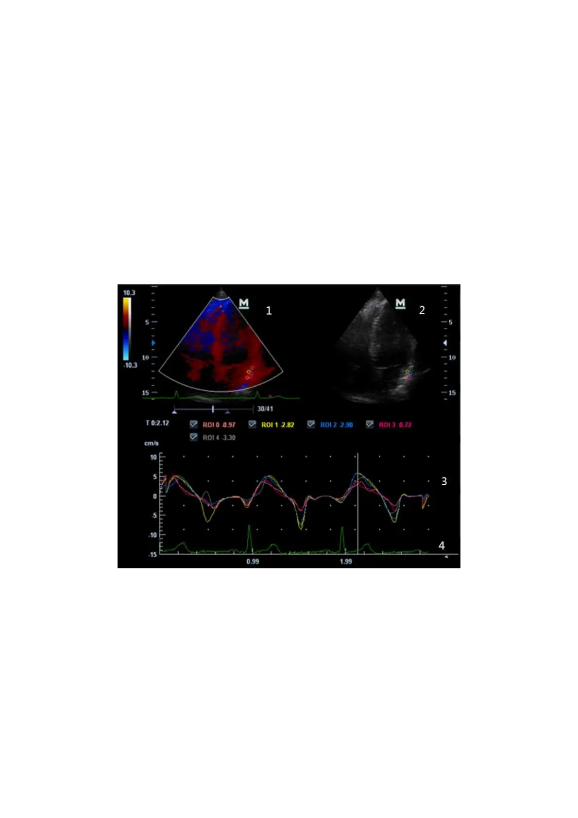
5.9.4.1 TDI QA Screen Description
1---TVI Cineloop window
Sample area: indicates sampling position of the analysis curve. The sample area is
color-coded, 8 (maximum) sample areas can be indicated.
2---B Cineloop window
Tips:
Images in the TVI cineloop window and B cineloop window are the frozen image
of the same moment; roll the trackball to review the images in the two cineloop
windows.
Sample areas are linked in the TVI cineloop window and B cineloop window.
3---Displays analysis curve
Y axis represents the velocity (unit: cm/s), while X axis represents the time (unit:
s).