Special Imaging Modes
11-2
11.1.1 Overview
Ultrasound data based on three-dimensional imaging methods can be used to image any
structure where a view can’t be achieved by standard 2D-mode to improve understanding
of complex structures.
This system supports Smart3D, Static 3D and 4D imaging modes. 4D provides
continuous, high volume acquisition of 3D images. 4D adds the dimension of “movement”
to a 3D image by providing continuous, real-time displays.
11.1.1.1 Terms
z Volume: a three-dimensional content.
z Volume data: the image data set of a 3D object rendered from a 2D image sequence.
z 3D image: the image displayed to represent the volume data.
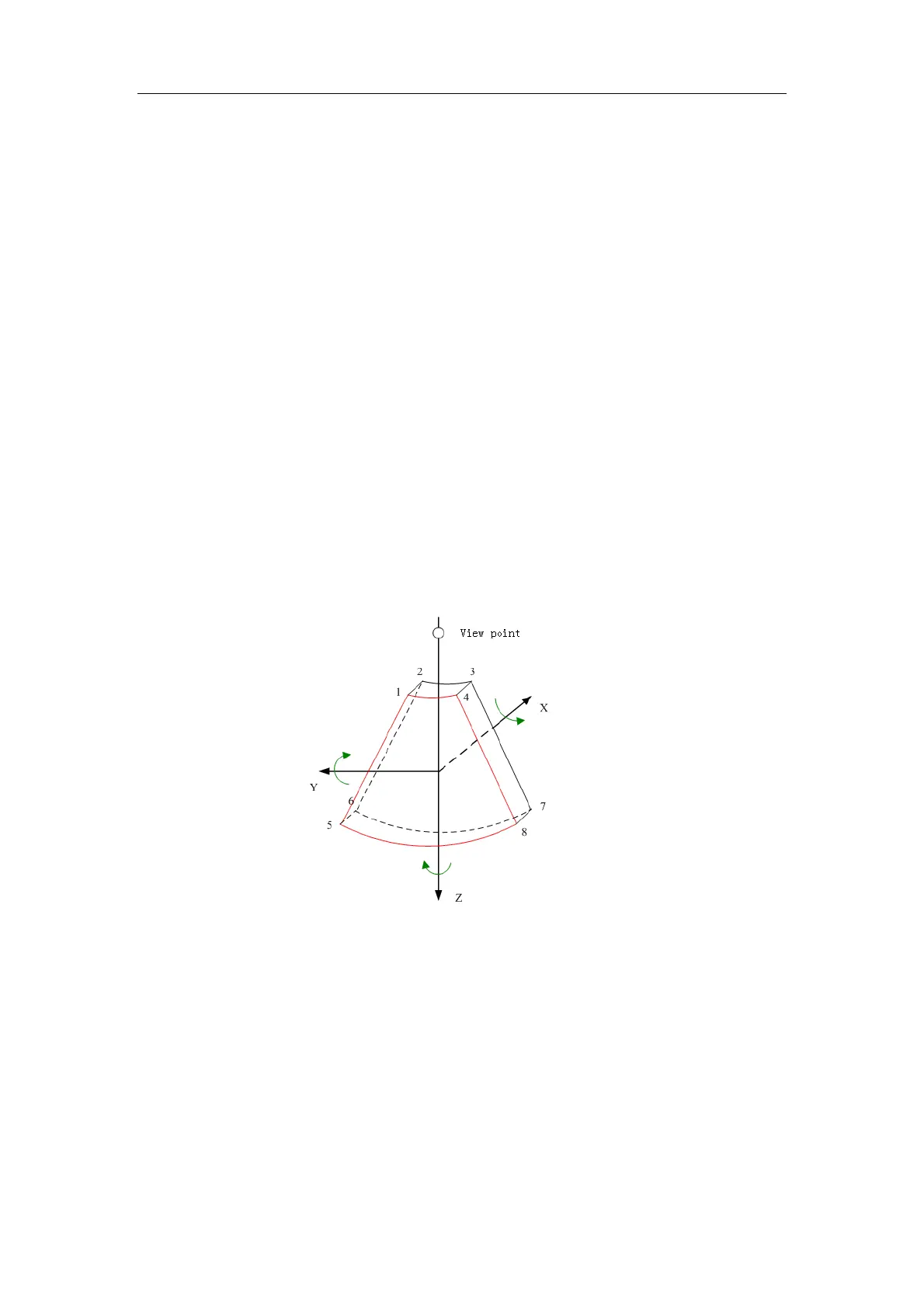
z View point: a position for viewing volume data, to obtain the 3D image.
z Sectional plane: tangent planes of the 3D image that obtained by algorithm. Of which,
XY-paralleled plane is C-plane, XZ-paralleled plane is B-plane, and YZ-paralleled plane is
A-plane. YZ-plane is the B scanning plane; the probe is moved along the X-axis.
z ROI (Region of Interest): a volume box used to determine the height and width of scanning
volume.
z VOI (Volume of Interest): a volume box used to determine the area of a sectional plane for
3D imaging.