System Options: Energy Dispersive X-ray (EDX) Analysis
C O N F I D E N T I A L – FEI Limited Rights Data 7-13
Energy Dispersive X-ray (EDX) Analysis
The EDX (sometimes referred to also as EDS analysis) is a technique used for identifying the elemental composition
of the specimen, or an area of interest thereof. It works as an integrated feature of a scanning electron microscope
(SEM), and cannot operate on its own without the latter.
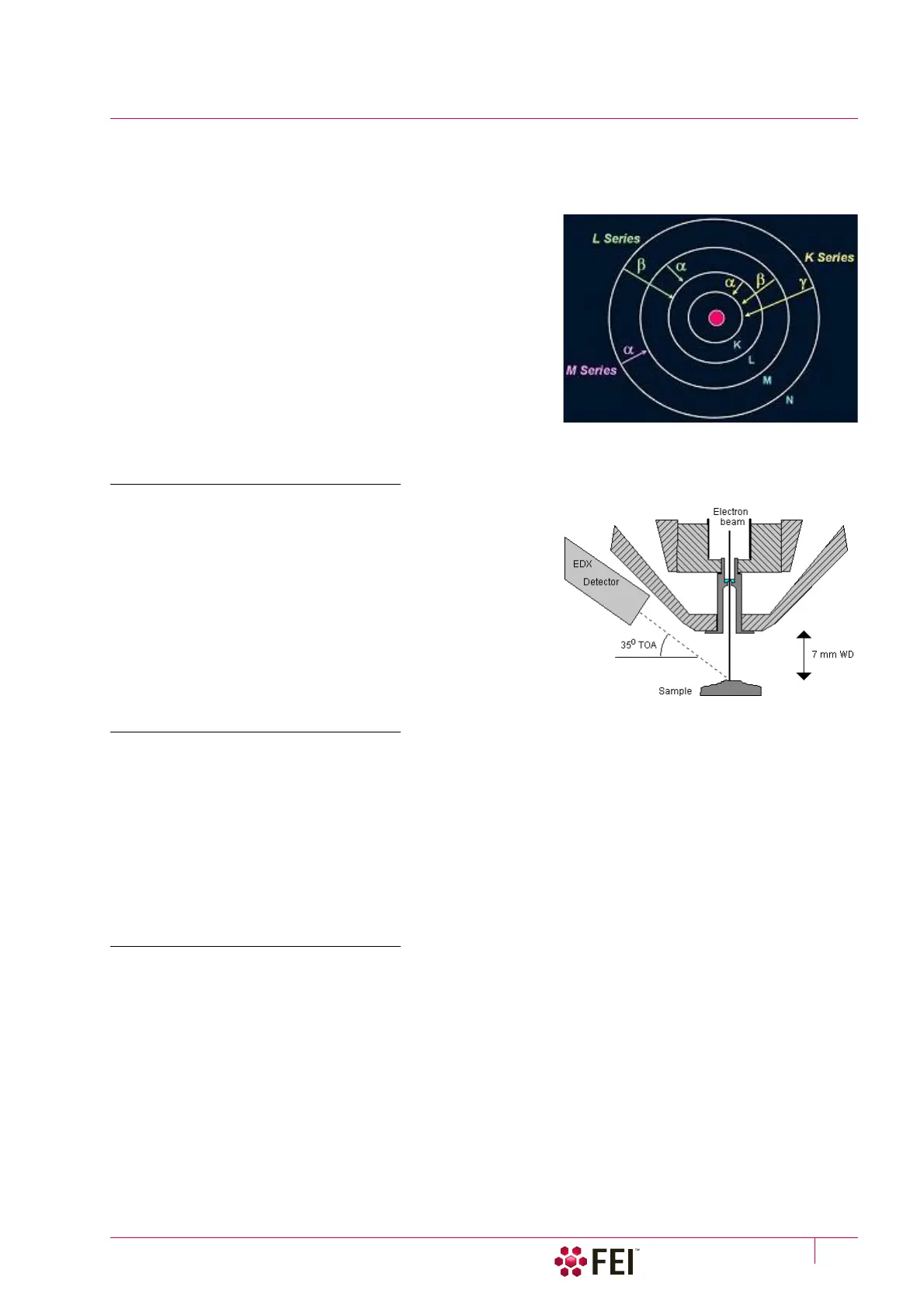
The specimen is bombarded with an electron beam inside the
microscope column. These electrons collide with the specimen
atoms' own electrons, knocking some of them off in the process.
Positions vacated by ejected inner shell electrons are occupied by
a higher-energy electron from an outer shell, while giving up
some of its energy by emitting an X-ray. The amount of energy
released depends on which shell it is transferring from / to. The
atom of every element releases X-rays with unique amount of
energy, identifying it.
The output of an EDX analysis is an EDX spectrum, which is just a
plot of how frequently an X-ray is received for each energy level.
The higher a peak in a spectrum, the more concentrated the
element is in the specimen.
High Vacuum
HiVac operation gives the most accurate X-ray results, but the
sample must be electrically conductive.
Low Vacuum EDX Analysis
X-ray analysis in LoVac mode is possible in combination either with the standard LVD detector, or with optional
GAD detector. The GAD is recommended to achieve the best signal-to-noise ratio, especially when using lower
accelerating values, because the long GAD cone minimizes the primary beam path and therefore its dispersion in
the gaseous environment of the chamber. On the other hand, the LVD detector offers the largest field of view for
the LoVac operation. The CBS is compatible with EDX.
The X-ray analysis should be performed at the lowest possible gas pressure to minimize the interaction of electrons
with the chamber gas. Normally, it is performed with a relatively high beam current so that there is enough signal
for a good LVD image even at very low gas pressures.
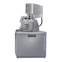
STEM EDX Analysis
Set the sample surface to 7 mm WD.
Select the area of interest in the STEM mode and perform X-ray analysis, mapping or line scans as appropriate.
Because the samples are not bulk in nature the beam spread normally associated with SEM samples is greatly
reduced and therefore higher spatial resolution can be obtained with the STEM detector. This also provides less
background in the spectrum and allows better separation of peaks as well as more accurate lower count rate
mapping. The high voltage chosen for the analysis still depends mainly on the composition of the sample.