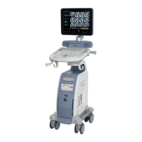
GE HEALTHCAREDRAFT VOLUSON® P8 / VOLUSON® P6
DIRECTION 5459672-100, R
EVISION 6 DRAFT (JANUARY 17, 2013) PROPRIETARY SERVICE MANUAL
Chapter 4 - Functional Checks 4-9
3
Focus Depth
To select the depth position of the actual focus zone(s). Arrows at the left
edge of the 2D-Image mark the active focal zone(s) by their depth position.
4
Depth
Adjusts the penetration depth range of the ultrasound image for the region
of interest.
The number of image lines and the frame rate are automatically optimized.
5
Multi Format (Dual, Quad)
Press this keys to change the display Mode from Single to DUAL or QUAD
display mode.
Press the SINGLE
format key or the 2D MODE key to change from Dual or
Quad to Single display.
6
2D Automatic Optimization
Pressing the AUTO key causes automatic optimization of the gray scale to
enhance the contrast resolution.
Pressing again: optimization will be updated and remain active.
Press the AUTO
key twice to switch off the Automatic Optimization in 2D.
7
TGCSlider Control
The °×TGC slide controls°± vary the gain in certain depths of the 2D image
to allow an exact compensation for the attenuation of the echoes over time
(depth).
TGC slide controls to selectively adjust the gain (brightness) in depth.
Slide a slide control to the left to decrease the gain in the corresponding
specific 2D depth.
Slide a slide control to the right to increase the gain in the corresponding
specific 2D depth.
8
XBEAM CRI
(CrossBeam
Compound Resolution Imaging)
Pulses are transmitted not only perpendicularly to the acoustic window, but
also in oblique directions. The advantages of XBeam CRI are enhanced
contrast resolution with better tissue differentiation and clear organ borders.
9
SRI
(Speckle Reduction Imaging)
Speckle Reduction Imaging is a smoothing type filter to reduce speckle in the
ultrasound image.
10
Receiver Frequency Range
The “Frequency range” function allows for the fast adjustment of high
resolution/lower penetration, mid resolution/mid penetration, or lower
resolution/ high penetration for the 2D image. From the transducer’s
broadband signal a certain start frequency and start bandwidth is extracted
and then continuously changed over depth. Every transducer has a set of
three fixed receive settings which are easily controlled by switching the
[Frequency] key.
11
Image Orientation
Use the LEFT/RIGHT respectively the UP/DOWN keys on the Voluson® P8
/ Voluson® P6 to alternate the image orientation.
12
ANGLE
Use this control to select a part of interest of the 2D image. The advantage
of the decreased field-of-view is an increased 2D frame rate due to the
smaller sector width.
13
ß-VIEW
This function allows the adjustment of the Volume O-Axis position of 3D
probes in 2D Mode. The green line in the displayed symbol indicates the
position of the acoustic block.
Table 4-3 2D Mode Functions
Step Task Expected Results