Appendix for Lorrca® MaxSis
Page 202 Lorrca Maxsis User Manual
Version 5.04 MRN-231-EN
RBC membrane stability
Using the Ektacytometer, Chasis et al. found that the continuous high shear stress slowly changes
the mechanical properties of resealed erythrocyte membranes. (Reference 11
7
) This causes the
cell membranes to fragment progressively. The process can be accelerated by further increasing
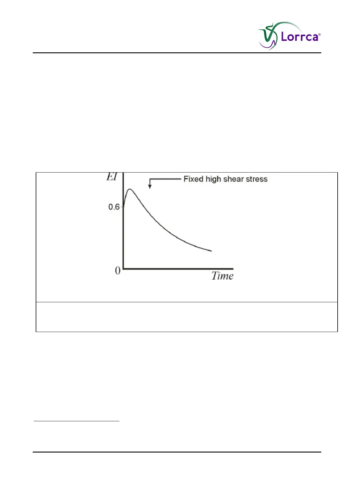
the applied shear stress. The diffraction pattern gradually looses its ellipticity, and hence EI
decreases, under continued high shear stress (see Figure 3). The decrease in EI can be explained
by the appearance of randomly oriented cell fragments which add a circular component to the –
initially- elliptical diffraction pattern. A similar effect was observed, using the LORCA, in mixtures of
rigid and normal deforming RBC, both in the experimental (REF) and clinical situation, e.g. in the
blood from patients with sickle cell disease (REF). However, for comparison of results obtained by
the Ektacytometer with those of the LORCA it should be kept in mind that the Ektacytometer uses
a different method to analyse the obtained diffraction pattern, i.e. 4 diodes detecting light intensity,
in contrast to LORCA`s modern digital image analysis.
Figure 3: Fixed high shear stress
Figure 3. A stability test (References (on page 215)
11
) is measured as the
elongation index (EI) vs. time while resealed erythrocyte membranes are
subject to a continuous high shear stress.
7
Chasis J.A., Mohandas N., Erythocyte membrane deformability and stability: two distinct membrane
properties that are independently regulated by skeletal protein associations, J. Cell Biol., vol. 103:(2), pp.
343-350, 1986.