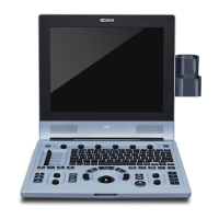
U60 Diagnostic Ultrasound System User Manual System Control
- 40 -
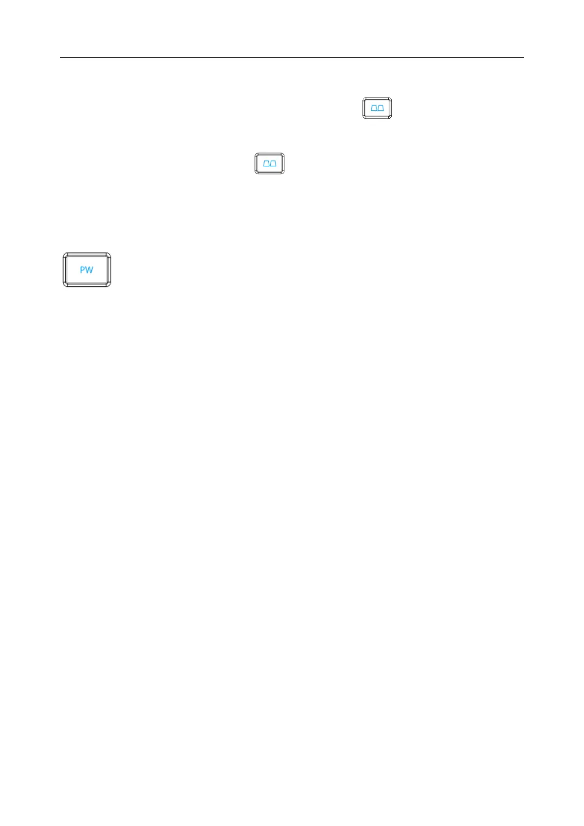
Dual-split mode of B+Color/PDI:
In dual-split mode of 2B, press Color/PDI, and then press to enter dual-split mode of
B+Color/PDI, the two windows are in B+Color/PDI mode.
In single B+Color/PDI mode, press to enter dual-split mode of B+Color/PDI, the two
windows are in B+Color/PDI mode.
In dual-split mode of B+Color/PDI, press Color/PDI, the currently active window exits color
mode and enters 2B mode.
Pulsed-Wave Doppler mode Display Control
In B mode, press this key to display the sample line, and press this key again to enter B+PW
mode; in B+PW mode, press this key to exit PW mode and enter B mode.
A pulsed-wave Doppler (PW) scan produces a series of pulses used to study the motion of blood
flow in a small region along a desired scan line, called the sample volume.
The X-axis of the graph represents time, and the Y-axis represents Doppler frequency shift. The
shift in frequency between successive ultrasound pulses, caused mainly by moving red blood
cells, can be converted into velocity and flow if an appropriate angle between the insonating
beam and blood flow is known.
Shades of gray in the spectral display represent the strength of the signal. The thickness of the
spectral signal is indicative of laminar or turbulent flow (laminar flow typically shows a narrow
band of blood flow information).
Pulsed-Wave Doppler mode and B mode are shown together in a mixed mode display. This
combination lets you monitor the exact location of the sample volume on the B image in the B
Image Display window, while acquiring Pulsed-Wave Doppler data in the Time Series window.
Operations:
In the B scan, the long line lets you adjust the sample line position, the two parallel lines (that
look like =) let you adjust the sample volume (SV) size and depth, and the line that crosses them
lets you adjust the correction angle (PW angle).